

Validation of a Novel Semiautomated Segmentation Method for MRI Detection of Cartilage-Related Bone Marrow Lesions
- PMID: 26069564
- PMCID: PMC4297059
- DOI: 10.1177/1947603510376819
Validation of a Novel Semiautomated Segmentation Method for MRI Detection of Cartilage-Related Bone Marrow Lesions
Abstract
Objective: To determine the relationship of bone marrow lesions (BMLs) with phenomena such as clinical symptoms, histological subchondral bone damage, and development of osteoarthritis, a reliable and reproducible method to localize and quantify BMLs accurately is indispensable. Therefore, the goal of the current study was to develop and validate a novel semiautomated segmentation method based on the KNN classification technique on T2-weighted (T2w) SPIR and proton density-weighted (PDw) magnetic resonance images (MRIs), as this would provide an accurate, reliable, and reproducible tool.
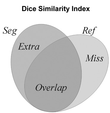
Materials and methods: Twenty PDw and T2w SPIR MRIs were selected and manually segmented as a learning set for the software system. The manual segmentations were considered the gold standard. Automated segmentation based on the KNN classification technique was carried out on the same MRIs. To determine the accuracy and validity of the system, the automated segmentations were compared to the gold standard using the Dice Similarity Index (DSI).
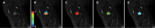
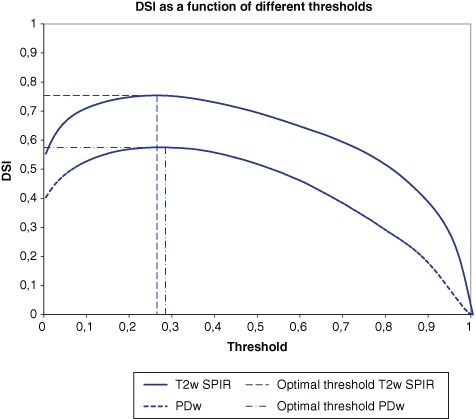
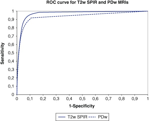
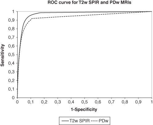
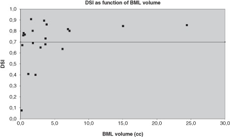
Results: The KNN classification system resulted both visually and statistically in an accurate segmentation of BMLs on T2w SPIR MRIs with an excellent mean optimal DSI of 0.702 (±0.202; range, 0.409-0.908). Elimination of specific areas smaller than 10 voxels improved the accuracy. The accuracy was independent of BML size. The segmentation of BMLs on PDw MRIs was less reliable with a mean optimal DSI of 0.536 (±0.156).
Conclusion: Although the applicability of this method is limited on PDw MRIs, the KNN classification system provides an accurate, reliable, and reproducible tool for semiautomated segmentation of BMLs in T2w SPIR MRIs of the knee.
Keywords: KNN classification; MRI; bone marrow edema; bone marrow lesions; cartilage; quantitative method; segmentation.
Conflict of interest statement
Figures
References
-
- Mink JH, Deutsch AL. Occult cartilage and bone injuries of the knee: detection, classification, and assessment with MR imaging. Radiology. 1989;170:823-9. - PubMed
-
- Hofmann S, Kramer J, Breitenseher M, Pietsch M, Aigner N. [Bone marrow edema in the knee. Differential diagnosis and therapeutic possibilities]. Orthopade. 2006;35:463-75. - PubMed
-
- Felson DT, McLaughlin S, Goggins J, LaValley MP, Gale ME, Totterman S, et al. Bone marrow edema and its relation to progression of knee osteoarthritis. Ann Intern Med. 2003;139:330-6. - PubMed
-
- Felson DT, Niu J, Guermazi A, Roemer F, Aliabadi P, Clancy M, et al. Correlation of the development of knee pain with enlarging bone marrow lesions on magnetic resonance imaging. Arthritis Rheum. 2007;56:2986-92. - PubMed
-
- Hunter DJ, Zhang Y, Niu J, Goggins J, Amin S, LaValley MP, et al. Increase in bone marrow lesions associated with cartilage loss: a longitudinal magnetic resonance imaging study of knee osteoarthritis. Arthritis Rheum. 2006;54:1529-35. - PubMed
LinkOut - more resources
Full Text Sources
