. 2011 Oct;2(4):327-36.
doi: 10.1177/1947603511400726.

The Treatment of Osteochondral Lesions of the Talus with Autologous Osteochondral Transplantation and Bone Marrow Aspirate Concentrate: Surgical Technique

Affiliations

The Treatment of Osteochondral Lesions of the Talus with Autologous Osteochondral Transplantation and Bone Marrow Aspirate Concentrate: Surgical Technique

John G Kennedy et al. Cartilage. 2011 Oct.

Abstract

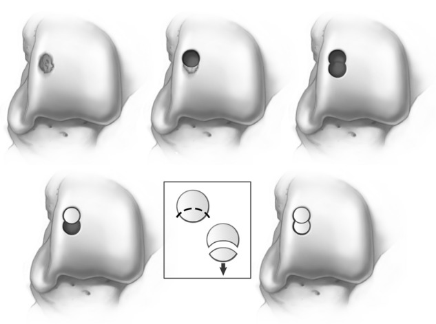
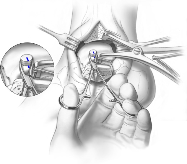
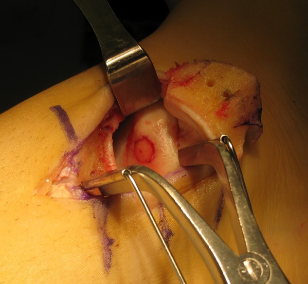
Objective: To present the functional results after autologous osteochondral transplantation with bone marrow aspirate concentrate in 72 patients, while placing an emphasis on the surgical technique.

Methods: Between 2005 and 2009, 72 patients underwent autologous osteochondral transplantation under the care of the senior author. The mean patient age at the time of surgery was 34.19 years (range, 16-85 years). All patients were followed for a minimum of 1 year after surgery. The mean follow-up time was 28.02 months (range, 12-64 months). Patient-reported outcome measures were taken preoperatively and at final follow-up using the Foot and Ankle Outcome Score (FAOS) and Short Form-12 (SF-12) general health questionnaire. Identical questionnaires were used in all instances.

Results: The mean FAOS scores improved from 52.67 points preoperatively to 86.19 points postoperatively (range, 71-100 points). The mean SF-12 scores also improved from 59.40 points preoperatively to 88.63 points postoperatively (range, 52-98 points). Three patients reported donor site knee pain after surgery, and one patient required the decompression of a cyst that developed beneath the graft site approximately 2 years after the index procedure.

Conclusion: Autologous osteochondral transplantation is a reproducible and primary treatment strategy for large osteochondral lesions of the talus.

Keywords: ankle < joint involved; articular cartilage < tissue; cartilage repair < repair; cartilage transplantation < grafts; sports injury < diagnosis.


Conflict of interest statement

Declaration of Conflicting Interests: The authors declared no potential conflicts of interest with respect to the authorship and/or publication of this article.

Figures

Figure 1.
Figure 1.
A provisional K-wire is drilled, exiting at the malleolar colliculus, to fluoroscopically visualize the medial malleolar osteotomy. A chevron-type cut is then made in the medial malleolus. Illustrations copyright of and reproduced with permission from JG Kennedy MD. Reproduction without express written consent is prohibited.
Figure 2.
Figure 2.
Two parallel fixation holes are predrilled in the medial malleolus prior to making the osteotomy cut Illustrations copyright of and reproduced with permission from JG Kennedy MD. Reproduction without express written consent is prohibited.
Figure 3.
Figure 3.
A modified retractor is utilized to allow adequate visualization and access to the medial aspect of the talus. Illustrations copyright of and reproduced with permission from JG Kennedy MD. Reproduction without express written consent is prohibited.
Figure 4.
Figure 4.
(A) A lateral tibial trapezoidal osteotomy can be made to provide access to all but the most posterior lesions, thereby avoiding fibular takedown. (B) A single predrilled 4-mm titanium screw is used as final fixation for the osteotomy Illustrations copyright of and reproduced with permission from JG Kennedy MD. Reproduction without express written consent is prohibited.
Figure 5.
Figure 5.
A half-moon or figure-of-8 graft configuration is used to prevent that empty space that is evident when 2 cylindrical grafts are placed adjacent to one another Illustrations copyright of and reproduced with permission from JG Kennedy MD. Reproduction without express written consent is prohibited.
Figure 6.
Figure 6.
Overdrilling and shaping the graft in a “bullet” fashion may prevent incongruency in the graft surface throughout the maturation process.
Figure 7.
Figure 7.
The lateral nonweightbearing aspect of the femoral condyle can be utilized for harvesting a donor plug Illustrations copyright of and reproduced with permission from JG Kennedy MD. Reproduction without express written consent is prohibited.
Figure 8.
Figure 8.
An ongoing biomechanical study at the authors’ institution to characterize the contact pressure alteration and restoration following the autologous osteochondral transplantation procedure demonstrates that relative to the intact ankle (A), creating an osteochondral lesion significantly alters contact pressure about the ankle joint such that the pressure is shifted centrally when loaded (B). Placement of the autologous osteochondral transplantation graft into the site of the defect (C) restores loading to intact levels.
Figure 9.
Figure 9.
A mosquito snap is used to rotate the graft into the most congruent position possible Illustrations copyright of and reproduced with permission from JG Kennedy MD. Reproduction without express written consent is prohibited.
Figure 10.
Figure 10.
The graft is tapped into final position only once perfect congruency is achieved.
Figure 11.
Figure 11.
Following graft placement, the osteotomy is fixated with titanium screws using the predrilled holes. The utilization of 2 medial malleolar screws at the osteotomy site has the potential to shift postoperatively (A). The current authors have modified this technique with a third transverse screw to prevent this potential shift in fixation (B).
Figure 12.
Figure 12.
Coronal quantitative T2-mapping assessment of an osteochondral autograft transplanted into the medial aspect of the talar dome demonstrates restoration of the radius of curvature and color stratification similar to that of native cartilage.

References

    1. Saxena A, Eakin C. Articular talar injuries in athletes: results of microfracture and autogenous bone graft. Am J Sports Med. 2007;35(10):1680-7. - PubMed
    1. Karlsson J, Sancone M. Management of acute ligament injuries of the ankle. Foot Ankle Clin. 2006;11(3):521-30. - PubMed
    1. Kannus P, Renstrom P. Treatment for acute tears of the lateral ligaments of the ankle: operation, cast, or early controlled mobilization. J Bone Joint Surg Am. 1991;73(2):305-12. - PubMed
    1. Lynch SA, Renström PA. Treatment of acute lateral ankle ligament rupture in the athlete conservative versus surgical treatment. Sports Med. 1999;27(1):61-71. - PubMed
    1. Chuckpaiwong B, Berkson EM, Theodore GH. Microfracture for osteochondral lesions of the ankle: outcome analysis and outcome predictors of 105 cases. Arthroscopy. 2008;24: 106-12. - PubMed

LinkOut - more resources