

Comparison of Three Methods to Quantify Repair Cartilage Collagen Orientation
- PMID: 26069654
- PMCID: PMC4297104
- DOI: 10.1177/1947603512461440
Comparison of Three Methods to Quantify Repair Cartilage Collagen Orientation
Abstract
Objective: The aim of this study was to determine if the noninvasive or minimally invasive and nondestructive imaging techniques of quantitative T2-mapping or multiphoton microscopy (MPM) respectively, could detect differences in cartilage collagen orientation similar to polarized light microscopy (PLM). It was hypothesized that MRI, MPM, and PLM would all detect quantitative differences between repair and normal cartilage tissue.
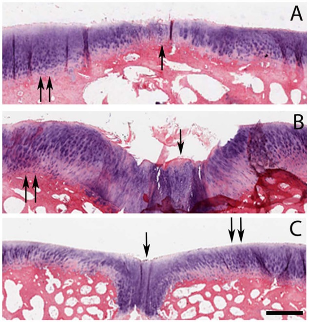
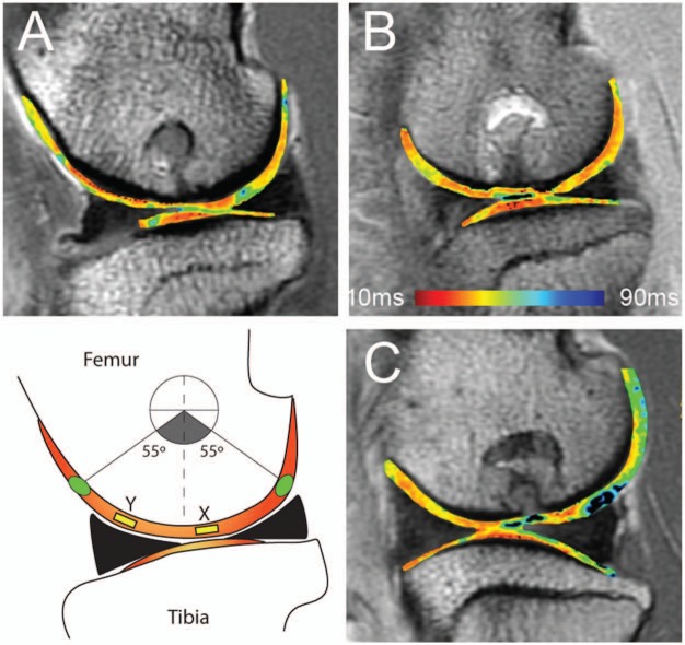
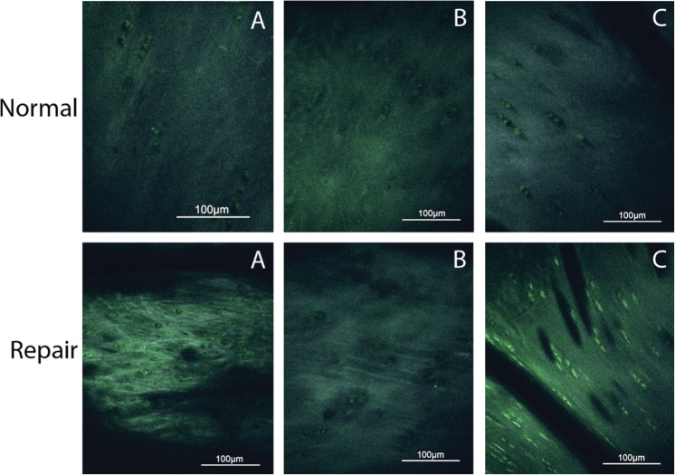
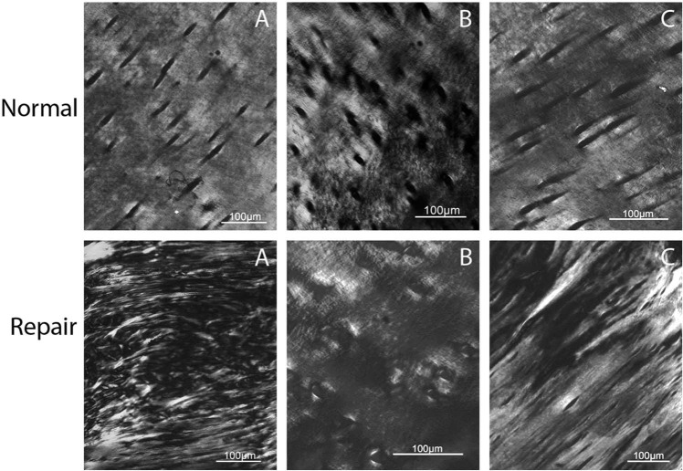
Methods: Osteochondral defects in the medial femoral condyle were created and repaired in 5 mature goats. Postmortem, MRI with T2-mapping and histology were performed. T2 maps were generated and a mean T2 value was calculated for each region of interest. Histologic slides were assessed using MPM with measurements of autocorrelation ellipticity, and by PLM with application of a validated scoring method. Collagen orientation using each of the 3 modalities (T2-mapping, MPM, and PLM) was measured in the center of the repair tissue and compared to remote, normal cartilage.
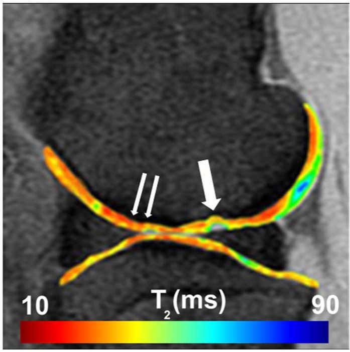
Results: MRI, MPM, and PLM were able to detect a significant difference between repair and normal cartilage (n = 5). The average T2 value was longer for repair tissue (41.43 ± 9.81 ms) compared with normal cartilage (27.12 ± 14.22 ms; P = 0.04); MPM autocorrelation ellipticity was higher in fibrous tissue (3.75 ± 1.17) compared with normal cartilage (2.24 ± 0.51; P = 0.01); the average PLM score for repair tissue was lower (1.6 ± 1.02) than the score for remote normal cartilage (4.4 ± 0.42; P = 0.002). The strongest correlation among the methods was between MRI and PLM (r = -0.76; P = 0.01), followed by MPM and PLM (r = -0.58; P = 0.08), with the weakest correlation shown between MRI and MPM (r = 0.35; P = 0.31).
Conclusion: All 3 imaging methods quantitatively measured differences in collagen orientation between repair and normal cartilage, but at very different levels of resolution. PLM is destructive to tissue and requires euthanasia, but because MPM can be used arthroscopically, both T2-mapping and MPM can be performed in vivo, offering nondestructive means to assess collagen orientation that could be used to obtain longitudinal data in cartilage repair studies.
Keywords: articular cartilage; cartilage repair; outcome measures; tissue.
Conflict of interest statement
Ethical Approval: This study was approved by our institutional review boards.
Figures
References
-
- Silver FH, Bradica G, Tria A. Relationship among biomechanical, biochemical, and cellular changes associated with arthritis. Crit Rev Biomed Eng. 2001;24(4):373-91. - PubMed
-
- Silver FH, Glasgold AI. Cartilage wound healing. An overview. Otolaryngol Clin North Am. 1995;28(5):847-64. - PubMed
-
- Weiss C, Rosenberg L, Helfet AJ. An ultrastructural study of normal adult human articular cartilage. J Bone Joint Surg. 1968;50(4):663-74. - PubMed
-
- Jeffery A, Blunn G, Archer C, Bentley G. Three-dimensional collagen architecture in bovine articular cartilage. J Bone Joint Surg Br. 1991;73:795-801. - PubMed
-
- Hunziker EB, Michel M, Studer D. Ultrastructure of adult human articular cartilage matrix after cryotechnical processing. Microsc Res Tech. 1997;37(4):271-84. - PubMed
Grants and funding
LinkOut - more resources
Full Text Sources
