. 2012 Jun;5(3):195-202.
doi: 10.1093/ckj/sfs029. Epub 2012 Mar 28.

Aquaporin-2: new mutations responsible for autosomal-recessive nephrogenic diabetes insipidus-update and epidemiology

Affiliations

Aquaporin-2: new mutations responsible for autosomal-recessive nephrogenic diabetes insipidus-update and epidemiology

Daniel G Bichet et al. Clin Kidney J. 2012 Jun.

Abstract

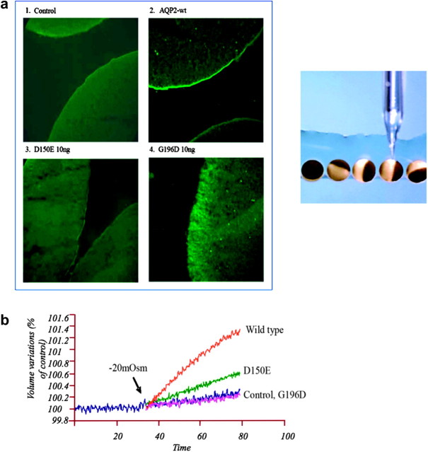
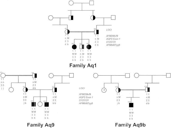
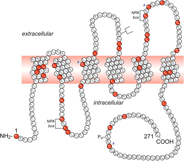
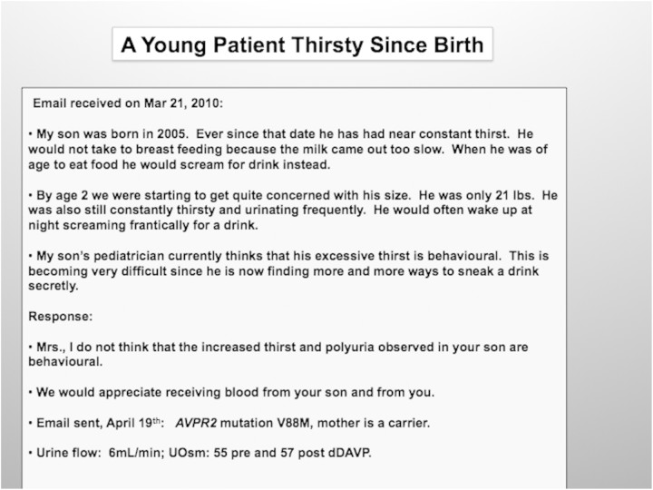
It is clinically useful to distinguish between two types of hereditary nephrogenic diabetes insipidus (NDI): a 'pure' type characterized by loss of water only and a complex type characterized by loss of water and ions. Patients with congenital NDI bearing mutations in the vasopressin 2 receptor gene, AVPR2, or in the aquaporin-2 gene, AQP2, have a pure NDI phenotype with loss of water but normal conservation of sodium, potassium, chloride and calcium. Patients with hereditary hypokalemic salt-losing tubulopathies have a complex phenotype with loss of water and ions. They have polyhydramnios, hypercalciuria and hypo- or isosthenuria and were found to bear KCNJ1 (ROMK) and SLC12A1 (NKCC2) mutations. Patients with polyhydramnios, profound polyuria, hyponatremia, hypochloremia, metabolic alkalosis and sensorineural deafness were found to bear BSND mutations. These clinical phenotypes demonstrate the critical importance of the proteins ROMK, NKCC2 and Barttin to transfer NaCl in the medullary interstitium and thereby to generate, together with urea, a hypertonic milieu. This editorial describes two new developments: (i) the genomic information provided by the sequencing of the AQP2 gene is key to the routine care of these patients, and, as in other genetic diseases, reduces health costs and provides psychological benefits to patients and families and (ii) the expression of AQP2 mutants in Xenopus oocytes and in polarized renal tubular cells recapitulates the clinical phenotypes and reveals a continuum from severe loss of function with urinary osmolalities <150 mOsm/kg H2O to milder defects with urine osmolalities >200 mOsm/kg H2O.

Keywords: aquaporin-2 mutations; autosomal-dominant and -recessive nephrogenic diabetes insipidus; genetic testing; hereditary polyuric states.


Figures

Fig. 1.
Fig. 1.
(a) Immunofluorescence of AQP2 expressed in oocytes. Oocytes were not injected (1 control) or injected with either AQP2-wt (2, 1 ng), AQP2-D150E (3, 10 ng) or AQP2-G196D (4, 10 ng) messenger RNAs and incubated for 3 days prior to assay. Oocytes were immunostained and visualized with antibodies to AQP2. The injection process is represented in the right of the figure [21]. (b) Determination of water permeabilities (Pf) of wild-type (WT) AQP2 and mutants expressed in Xenopus oocytes. Oocytes were injected with either AQP2-wt (1 ng), AQP2-D150E (10 ng) or AQP2-G196D (10 ng) messenger RNAs and incubated for 2 days prior to assay. Determination of water permeabilities was performed by evaluation of volume increase in oocytes as induced by a 20-mosmol/kg H2O hypotonic shock [21].
Fig. 2.
Fig. 2.
Pedigree of three Pakistani families referred to our laboratory each bearing the V71M AQP2 mutation (M). Family Aqp1 was first described by Langley [16]. Squares and circles represent male and female subjects, respectively, with unaffected individuals (open symbols), carriers (half-filled symbols) and affected individuals (solid symbol); n indicates normal allele. Haplotypes consist of markers that flank the AQP2 gene and that have been described previously [24]. The alleles bearing the individual mutations are identical suggesting a common ancestry.
Fig. 3.
Fig. 3.
A representation of the AQP2 protein and identification of 46 putative disease-causing AQP2 mutations. A monomer is represented with six transmembrane helices. The location of the PKA phosphorylation site (Pa) is indicated. The extracellular, transmembrane and cytoplasmic domains are defined according to Deen et al. [17]. Solid symbols indicate the location of the mutations (for references, view Table 1): M1I; L22V; V24A; L28P; G29S; A47V; Q57P; G64R; N68S; A70D; V71M; R85X; G100X; G100V; G100R; I107D; 369delC; T125M; T126M; A147T; D150E; V168M; G175R; G180S; C181W; P185A; R187C; R187H; A190T; G196D; W202C; G215C; S216P; S216F; K228E; R254Q; R254L; E258K and P262L. GenBank accession numbers—AQP2: AF147092, Exon 1; AF147093, Exons 2 through 4. NPA motifs and the N-glycosylation site are also indicated.
Fig. 4.
Fig. 4.
A young patient's thirsty since birth.
Fig. 5.
Fig. 5.
Schematic representation of the effect of vasopressin (AVP) to increase water permeability in the principal cells of the collecting duct. AVP is bound to the V2 receptor (a G-protein-linked receptor) on the basolateral membrane. The basic process of G-protein-coupled receptor signaling consists of three steps: a hepta-helical receptor that detects a ligand (in this case, AVP) in the extracellular milieu, a G-protein (Gα s) that dissociates into a subunits bound to guanosine triphosphate and bg subunits after interaction with the ligand-bound receptor and an effector (in this case, adenylyl cyclase) that interacts with dissociated G-protein subunits to generate small molecule second messengers. AVP activates adenylyl cyclase, increasing the intracellular concentration of cAMP. The topology of adenylyl cyclase is characterized by two tandem repeats of six hydrophobic transmembrane domains separated by a large cytoplasmic loop and terminates in a large intracellular tail. The dimeric structure (C1 and C2) of the catalytic domains is represented. Conversion of adenosine triphosphate (ATP) to cAMP takes place at the dimer interface. Two aspartate residues (in C1) coordinate two metal co-factors (Mg2+ or Mn2+ represented here as two small black circles), which enable the catalytic function of the enzyme. Adenosine is shown as an open circle and the three phosphate groups (ATP) are shown as smaller open circles. PKA is the target of the generated cAMP. The binding of cAMP to the regulatory subunits of PKA induces a conformational change, causing these subunits to dissociate from the catalytic subunits. These activated subunits (C) as shown here are anchored to an aquaporin-2 (AQP2)-containing endocytic vesicle via an A-kinase-anchoring protein. The local concentration and distribution of the cAMP gradient are limited by phosphodiesterases (PDEs). Cytoplasmic vesicles carrying the water channels (represented as homotetrameric complexes) are fused to the luminal membrane in response to AVP, thereby increasing the water permeability of this membrane. The dissociation of the A-kinase-anchoring protein from the endocytic vesicle is not represented. Microtubules and actin filaments are necessary for vesicle movement toward the membrane. When AVP is not available, AQP2 water channels are retrieved by an endocytic process, and water permeability returns to its original low rate. Aquaporin-3 (AQP3) and aquaporin-4 (AQP4) water channels are expressed constitutively at the basolateral membrane.

References

    1. Estevez R, Boettger T, Stein V, et al. Barttin is a Cl- channel beta-subunit crucial for renal Cl- reabsorption and inner ear K+ secretion. Nature. 2001;414:558–561. - PubMed
    1. Waldegger S, Jeck N, Barth P, et al. Barttin increases surface expression and changes current properties of ClC-K channels. Pflugers Arch. 2002;444:411–418. - PubMed
    1. Jeck N, Reinalter SC, Henne T, et al. Hypokalemic salt-losing tubulopathy with chronic renal failure and sensorineural deafness. Pediatrics. 2001;108:E5. - PubMed
    1. Birkenhager R, Otto E, Schurmann MJ, et al. Mutation of BSND causes Bartter syndrome with sensorineural deafness and kidney failure. Nat Genet. 2001;29:310–314. - PubMed
    1. Bettinelli A, Bianchetti MG, Girardin E, et al. Use of calcium excretion values to distinguish two forms of primary renal tubular hypokalemic alkalosis: Bartter and Gitelman syndromes. J Pediatr. 1992;120:38–43. - PubMed