

Reduced Number of Pigmented Neurons in the Substantia Nigra of Dystonia Patients? Findings from Extensive Neuropathologic, Immunohistochemistry, and Quantitative Analyses
- PMID: 26069855
- PMCID: PMC4458735
- DOI: 10.7916/D8T72G9G
Reduced Number of Pigmented Neurons in the Substantia Nigra of Dystonia Patients? Findings from Extensive Neuropathologic, Immunohistochemistry, and Quantitative Analyses
Abstract
Background: Dystonias (Dys) represent the third most common movement disorder after essential tremor (ET) and Parkinson's disease (PD). While some pathogenetic mechanisms and genetic causes of Dys have been identified, little is known about their neuropathologic features. Previous neuropathologic studies have reported generically defined neuronal loss in various cerebral regions of Dys brains, mostly in the basal ganglia (BG), and specifically in the substantia nigra (SN). Enlarged pigmented neurons in the SN of Dys patients with and without specific genetic mutations (e.g., GAG deletions in DYT1 dystonia) have also been described. Whether or not Dys brains are associated with decreased numbers or other morphometric changes of specific neuronal types is unknown and has never been addressed with quantitative methodologies.
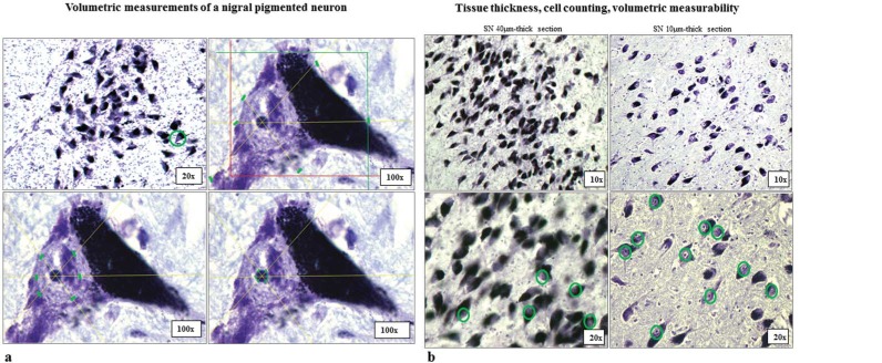
Methods: Quantitative immunohistochemistry protocols were used to estimate neuronal counts and volumes of nigral pigmented neurons in 13 SN of Dys patients and 13 SN of age-matched control subjects (C).
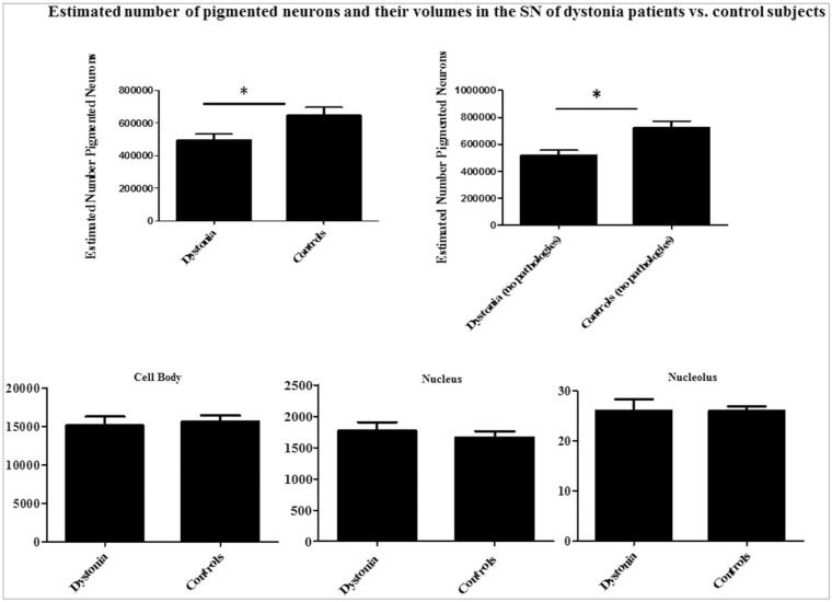
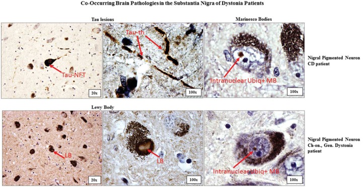
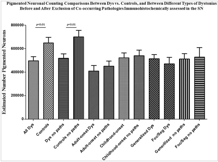
Results: We observed a significant reduction (∼20%) of pigmented neurons in the SN of Dys compared to C (p<0.01). Neither significant volumetric changes nor evident neurodegenerative signs were observed in the remaining pool of nigral pigmented neurons in Dys brains. These novel quantitative findings were confirmed after exclusion of possible co-occurring SN pathologies including Lewy pathology, tau-neurofibrillary tangles, β-amyloid deposits, ubiquitin (ubiq), and phosphorylated-TAR DNA-binding protein 43 (pTDP43)-positive inclusions.
Discussion: A reduced number of nigral pigmented neurons in the absence of evident neurodegenerative signs in Dys brains could indicate previously unconsidered pathogenetic mechanisms of Dys such as neurodevelopmental defects in the SN.
Keywords: Substantia nigra; neurodegenerative disorder; neurodevelopmental disorder; neuronal loss; neuronal reduction; pigmented neurons.
Conflict of interest statement
Figures
Similar articles
-
Hypertrophy of nigral neurons in Torsin1A deletion (DYT1) carriers manifesting dystonia.Parkinsonism Relat Disord. 2019 Jan;58:63-69. doi: 10.1016/j.parkreldis.2018.08.020. Epub 2018 Aug 31. Parkinsonism Relat Disord. 2019. PMID: 30193818
-
Parkinson disease and incidental Lewy body disease: Just a question of time?Neurology. 2015 Nov 10;85(19):1670-9. doi: 10.1212/WNL.0000000000002102. Epub 2015 Oct 14. Neurology. 2015. PMID: 26468408 Free PMC article.
-
Substantia nigra lesions in Alzheimer disease and normal aging.Alzheimer Dis Assoc Disord. 1995 Summer;9(2):61-7. doi: 10.1097/00002093-199509020-00001. Alzheimer Dis Assoc Disord. 1995. PMID: 7662324
-
Chapter 33: the history of movement disorders.Handb Clin Neurol. 2010;95:501-46. doi: 10.1016/S0072-9752(08)02133-7. Handb Clin Neurol. 2010. PMID: 19892136 Review.
-
Therapeutic potentials of plant iridoids in Alzheimer's and Parkinson's diseases: A review.Eur J Med Chem. 2019 May 1;169:185-199. doi: 10.1016/j.ejmech.2019.03.009. Epub 2019 Mar 8. Eur J Med Chem. 2019. PMID: 30877973 Review.
Cited by
-
Neuropathology of Dystonia.Tremor Other Hyperkinet Mov (N Y). 2019 Feb 25;9:569. doi: 10.7916/d8-j6sx-b156. eCollection 2019. Tremor Other Hyperkinet Mov (N Y). 2019. PMID: 30886764 Free PMC article. Review.
-
The Effectiveness of Transcranial Brain Stimulation in Improving Clinical Signs of Hyperkinetic Movement Disorders.Front Neurosci. 2016 Jan 7;9:486. doi: 10.3389/fnins.2015.00486. eCollection 2015. Front Neurosci. 2016. PMID: 26778947 Free PMC article. Review.
-
Possible Post-Traumatic Focal Dystonia Associated with Tau Pathology Localized to Putamen-Globus Pallidus.Mov Disord Clin Pract. 2018 Aug 8;5(5):492-498. doi: 10.1002/mdc3.12626. eCollection 2018 Sep-Oct. Mov Disord Clin Pract. 2018. PMID: 30637269 Free PMC article.
-
Motor network reorganization associated with rTMS-induced writing improvement in writer's cramp dystonia.Brain Stimul. 2025 Mar-Apr;18(2):198-210. doi: 10.1016/j.brs.2025.02.005. Epub 2025 Feb 7. Brain Stimul. 2025. PMID: 39924101
-
Is Generalized and Segmental Dystonia Accompanied by Impairments in the Dopaminergic System?Front Neurol. 2021 Nov 18;12:751434. doi: 10.3389/fneur.2021.751434. eCollection 2021. Front Neurol. 2021. PMID: 34867735 Free PMC article.
References
-
- Defazio G, Abbruzzese G, Livrea P, Berardelli A. Epidemiology of primary dystonia. Lancet Neurol. 2004;3:673–678. - PubMed
-
- Steeves TD, Day L, Dykeman J, Jette N, Pringsheim T. The prevalence of primary dystonia: a systematic review and meta-analysis. Mov Disord. 2012;27:1789–1796. - PubMed
-
- Marsden CD. Dystonia: the spectrum of the disease. Res Publ Assoc Res Nerv Ment Dis. 1976;55:351–367. - PubMed
LinkOut - more resources
Full Text Sources
Molecular Biology Databases
