

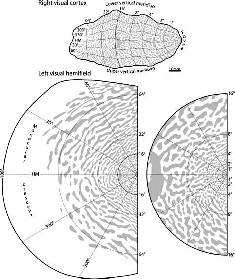
Refined Frequency Doubling Perimetry Analysis Reaffirms Central Nervous System Control of Chronic Glaucomatous Neurodegeneration
- PMID: 26069866
- PMCID: PMC4461216
- DOI: 10.1167/tvst.4.3.7
Refined Frequency Doubling Perimetry Analysis Reaffirms Central Nervous System Control of Chronic Glaucomatous Neurodegeneration
Abstract
Purpose: Refined analysis of frequency doubling perimetric data was performed to assess binocular visual field conservation in patients with comparable degrees of bilateral glaucomatous damage, to determine whether unilateral visual field loss is random, anatomically symmetric, or non-random in relation to the fellow eye.
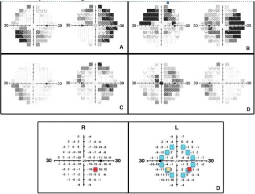
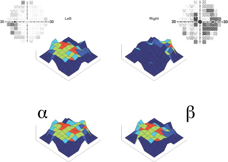
Methods: Case control study of 41 consecutive patients with bilaterally mild to severe glaucoma; each right eye visual field locus was paired with randomly-selected co-isopteric left eye loci, performing 690,000 (10,000 complete sets of 69 loci) such iterations per subject. The potential role of anatomic symmetry in bilateral visual field conservation was also assessed by pairing mirror-image loci of the right- and left-eye fields. The mean values of the random co-isopteric and the symmetric mirror pairings were compared with natural point-for-point pairings of the two eyes by paired t-test.
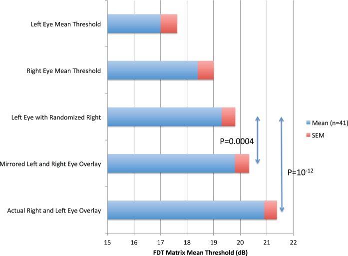
Results: Mean unilateral Matrix threshold across the entire 30-degree visual field were 17.0 dB left and 18.4 dB right (average 17.7). The better of the naturally paired concomitant loci yielded binocular equivalent mean bilateral Matrix threshold of 20.9 dB, 1.6 dB higher than the population mean of the 690,000 coisopteric pairings (t = -10.4; P < 10-12). Thus, a remarkable natural tendency for conservation of the binocular Matrix visual field was confirmed, far stronger than explicable by random chance. Symmetric pairings of precise mirror-image loci also produced values higher than random co-isopteric pairings (Δ 1.1 dB; t = -4.0; P = 0.0004).
Conclusions: Refined data analysis of paired Matrix visual fields confirms the existence of a natural optimization of binocular visual function in severe bilateral glaucoma via interlocking fields that could only be created by CNS involvement. The disparity of paired Matrix threshold values at mirror-image loci was also highly nonrandom and quantitatively inverse from the expected if anatomic symmetry factors were merely passively contributing systematically to the compensatory binocular Matrix effect.
Translational relevance: The paired eyes and brain are reaffirmed to function as a unified system in the progressive age-related neurodegenerative condition chronic open angle glaucoma, maximizing the binocular visual field. Given the extensive homology of this disorder with other age-related neurodegenerations, it is reasonable to assume that the brain will similarly resist simultaneous bilateral loss of paired functional zones in both hemispheres in diseases like Alzheimer's and Parkinson's disease. Glaucomatous eyes at all stages of the disease appear to provide a highly accessible paired-organ study model for developing therapeutics to optimize conservation of function in neurodegenerative disorders.
Keywords: frequency doubling; glaucoma; jigsaw effect; neurodegeneration; neuroprotection; perimetry; refined data analysis; visual fields.
Figures
Similar articles
-
Refined Data Analysis Provides Clinical Evidence for Central Nervous System Control of Chronic Glaucomatous Neurodegeneration.Transl Vis Sci Technol. 2014 May 6;3(3):1. doi: 10.1167/tvst.3.3.1. eCollection 2014 May. Transl Vis Sci Technol. 2014. PMID: 24932429 Free PMC article.
-
Performance of frequency-doubling technology perimetry in a population-based prevalence survey of glaucoma: the Tajimi study.Ophthalmology. 2007 Jan;114(1):27-32. doi: 10.1016/j.ophtha.2006.06.041. Epub 2006 Oct 27. Ophthalmology. 2007. PMID: 17070580
-
Humphrey matrix frequency doubling perimetry for detection of visual-field defects in open-angle glaucoma.Br J Ophthalmol. 2009 May;93(5):582-8. doi: 10.1136/bjo.2007.119909. Epub 2008 Jul 31. Br J Ophthalmol. 2009. PMID: 18669543
-
Visual function-specific perimetry for indirect comparison of different ganglion cell populations in glaucoma.Invest Ophthalmol Vis Sci. 2000 Jun;41(7):1783-90. Invest Ophthalmol Vis Sci. 2000. PMID: 10845599
-
Cognitive Dysfunctions in Glaucoma: An Overview of Morpho-Functional Mechanisms and the Impact on Higher-Order Visual Function.Front Aging Neurosci. 2021 Oct 6;13:747050. doi: 10.3389/fnagi.2021.747050. eCollection 2021. Front Aging Neurosci. 2021. PMID: 34690746 Free PMC article. Review.
Cited by
-
Mechanisms implicated in the contralateral effect in the central nervous system after unilateral injury: focus on the visual system.Neural Regen Res. 2021 Nov;16(11):2125-2131. doi: 10.4103/1673-5374.310670. Neural Regen Res. 2021. PMID: 33818483 Free PMC article. Review.
-
Neural Conduction Along Postretinal Visual Pathways in Glaucoma.Front Aging Neurosci. 2021 Aug 2;13:697425. doi: 10.3389/fnagi.2021.697425. eCollection 2021. Front Aging Neurosci. 2021. PMID: 34408643 Free PMC article.
-
Contributions of Brain Microstructures and Metabolism to Visual Field Loss Patterns in Glaucoma Using Archetypal and Information Gain Analyses.Invest Ophthalmol Vis Sci. 2024 Jul 1;65(8):15. doi: 10.1167/iovs.65.8.15. Invest Ophthalmol Vis Sci. 2024. PMID: 38975942 Free PMC article.
-
Widespread brain reorganization perturbs visuomotor coordination in early glaucoma.Sci Rep. 2019 Oct 2;9(1):14168. doi: 10.1038/s41598-019-50793-x. Sci Rep. 2019. PMID: 31578409 Free PMC article.
-
Cholinergic nervous system and glaucoma: From basic science to clinical applications.Prog Retin Eye Res. 2019 Sep;72:100767. doi: 10.1016/j.preteyeres.2019.06.003. Epub 2019 Jun 23. Prog Retin Eye Res. 2019. PMID: 31242454 Free PMC article. Review.
References
-
- Caprioli J. Automated perimetry in glaucoma. : Walsh TJ. Visual Fields: Examination and Interpretation. San Francisco, CA: American Academy of Ophthalmology; 1990: 71–105.
-
- Ferreira SM,, Lerner SF, Brunzini R,, Evelson PA, Llesuy SF. Oxidative stress markers in aqueous humor of glaucoma patients. Am J Ophthalmol. 2004; 137: 62–69. - PubMed
-
- Kumar DM, Agarwal N. Oxidative stress in glaucoma: a burden of evidence. J Glaucoma. 2007; 16: 334–343. - PubMed
LinkOut - more resources
Full Text Sources
Other Literature Sources
Miscellaneous
