Review
. 2015 Oct;72(19):3677-93.
doi: 10.1007/s00018-015-1948-5. Epub 2015 Jun 13.

Structure of potassium channels

Affiliations
Review

Structure of potassium channels

Qie Kuang et al. Cell Mol Life Sci. 2015 Oct.

Abstract

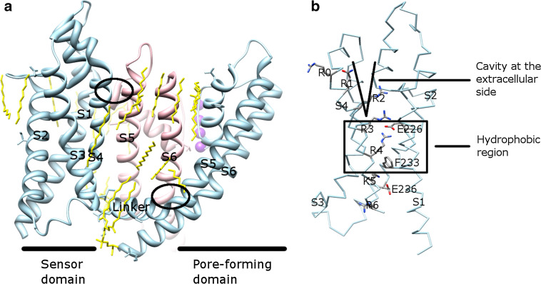
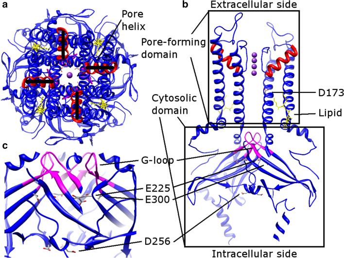
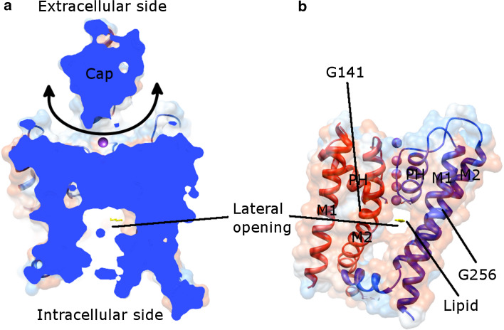
Potassium channels ubiquitously exist in nearly all kingdoms of life and perform diverse but important functions. Since the first atomic structure of a prokaryotic potassium channel (KcsA, a channel from Streptomyces lividans) was determined, tremendous progress has been made in understanding the mechanism of potassium channels and channels conducting other ions. In this review, we discuss the structure of various kinds of potassium channels, including the potassium channel with the pore-forming domain only (KcsA), voltage-gated, inwardly rectifying, tandem pore domain, and ligand-gated ones. The general properties shared by all potassium channels are introduced first, followed by specific features in each class. Our purpose is to help readers to grasp the basic concepts, to be familiar with the property of the different domains, and to understand the structure and function of the potassium channels better.

Keywords: Conductivity; Gating; RCK; Selectivity; Sensor domain.


Conflict of interest statement

No conflict of interest is declared.

Figures

Fig. 1
Fig. 1
The transmembrane part of KcsA. a The atomic structure of KcsA in the conductive state (PDB: 1K4C) viewed along the membrane plane. The pore-forming domain consists of the outer helix (magenta), loop regions (green), pore helix (blue), SF (yellow), and inner helix (orange). The conducted K+ ions are represented by purple balls with surrounding water molecules in red. EC is extracellular and IC is intracellular for short. The glycine hinge (Gly99) and the helical bundle are labeled. b, c The enlarged view of the boxed area in (a) containing the SF and the extracellular entryway. The K+ ions are in two configurations, either in S2 and S4 (b) or S1 and S3 (c) during conduction. The water molecules occupy the vacant ion positions in S1 and S3 (b) or in S2 and S4 (c). Other ions are located in the extracellular entryway (either S0 (b) or Sext (c)) and in the central cavity (Sc (a)). For clarity, only two monomers opposite to each other are shown. The amino acid sequence of the SF is labeled. All figures (Figs. 1, 2, 3, 4, 5, 6, 7, 8) in this paper were made using Chimera [130] and GNU Image Manipulation Program (GIMP)
Fig. 2
Fig. 2
Structural comparison of bacterial K+ channels: KcsA (PDB: 1K4C, cyan), KcsA (PDB: 3F5W [45], magenta), KvAP (PDB: 1ORQ, yellow), and MthK (PDB: 3LDC [24], blue). Except for KcsA in the closed conformation (cyan), the others are in open conformation. All structures are viewed from the intracellular side. The glycine hinges are in a similar position in these proteins. The outer helix, inner helix, and a queue of K+ ions are labeled
Fig. 3
Fig. 3
Activated, inactivated, and flipped SF structures of KcsA, viewed along the membrane plane. a Comparison of conductive (PDB: 1K4C, black) and nonconductive (PDB: 1K4D, 3F7V and 3F5W resemble each other and 3F7V is shown in magenta) structures. V76 and G77 are reorientated in the nonconductive state. b Comparison of conductive (PDB: 1K4C, black) and flipped (PDB: 2ATK [21] and 3OGC [22] are similar and 2ATK is shown in gray) structures. V76 and Y78 are reorientated in the flipped conformation. The S2 and S4 binding sites are labeled
Fig. 4
Fig. 4
The VSDs in channels. a Alignment of monomers of different channels, viewed from the extracellular side. When pore-forming domains are aligned, the VSDs adopt various orientations. The VSD (in an ellipse) is composed of the first four helices (S1–S4). The pore-forming domain (in a box) consists of S5 (corresponding to the outer helix in KcsA in Fig. 1) and S6 (corresponding to the inner helix in KcsA in Fig. 1). The pore helix is labeled as PH. b Alignment of published VSDs structures, viewed along the membrane plane. b Shows an enlarged side view, rotated 90° from (a). Different VSDs are compared: Kv1.2 (a Kv channel from Rattus norvegicus, PDB: 3LUT, light magenta), MlotiK1 (a non-voltage-gated K+ channel from Mesorhizobium loti, PDB: 3BEH, light brown), NavAb (a Nav channel from Arcobacter butzleri, PDB: 3RVY [131], light green), NavRh (a Nav channel from Rickettsiales sp. HIMB114, PDB: 4DXW [132], light orange), and TRPV1 (a transient receptor potential channel from Rattus norvegicus, PDB: 3J5P [133], light blue). Although the VSDs adopt different orientations in the channels (a), they show a substantial overlap when only these domains are compared (b). The VSDs in PDB: 3LUT, 3RVY, and 4DWX are aligned best. Two loop regions between S1–S2 and S3–S4 in PDB: 3LUT are omitted. The resembling structures (PDB: 2A79, 2R9R, 3RVZ [131], 3RW0 [131], 4EKW [134], 3J5R [37], and 3J5Q [37]) are not depicted. The VSD of KvAP is not shown either, since the solved structure is either distorted (PDB: 1ORQ) or resembles (PDB: 1ORS [31] and 2KYH [70]) the one in Kv1.2 (PDB: 3LUT)
Fig. 5
Fig. 5
Kv1.2–Kv2.1 chimera channel (PDB: 2R9R) and its VSD. a The entire channel. The linker in one subunit (light blue) locates below the pore-forming domain of another subunit (pink). Two interfaces are in ellipses. The lipids (yellow) surround the channel and fill into the empty space between the pore-forming domain and the VSD. Each individual TM is labeled. b The VSD structure. The positive residues, the counterbalanced negative residues, the hydrophobic region (in a box), and the cavity at the extracellular side are labeled. a and b are viewed along the membrane plane
Fig. 6
Fig. 6
Kir2.2 structure (PDB: 3SPI). a Extracellular view. The pore helices (red) are misaligned. b Side view, rotated 90° from (a). The pore-forming domain locates above the cytosolic domain. The G-loop (residues from 301 to 311, magenta), and conserved multiple ion binding sites (gray) are labeled. The PIP2 lipids (yellow) are located at the interface between two domains. c The extended ion conduction pathway in the cytosolic domain, an enlarged view of (b)
Fig. 7
Fig. 7
Human TWIK-1 structure (PDB: 3UKM) viewed along the membrane plane. a Cutaway view of the entire channel. The cap, a unique structure in K2P channels makes K+ ions (purple) coming laterally (indicated by a double end arrow). b Shows the lateral opening together with the channel model. Two adjacent subunits (red and blue) are shown. M1 (outer helix), M2 (inner helix), and PH (pore helix) are labeled, as well as the glycine hinges (G141 and G256). The structure presented here is in the down state. A lipid molecule (yellow) in the lateral opening is depicted. For clarity, the cap is omitted in (b) and only one lateral opening is displayed
Fig. 8
Fig. 8
RCK gating ring of KtrA (PDB: 2HMW [116]). a Is viewed down the fourfold axis from the extracellular side. The RCK monomers in the top layer (blue) and bottom layer (red) are connected through the alternative flexible and assembly interfaces, but the monomers in the same layer do not interact with each other. (b) Is the side view, rotated 90° from (a). One RCK monomer (red) interacts with two adjacent monomers in another layer (blue). The adenosine triphosphate ligands are shown in yellow

References

    1. Kuo MM, Haynes WJ, Loukin SH, Kung C, Saimi Y. Prokaryotic K(+) channels: from crystal structures to diversity. FEMS Microbiol Rev. 2005;29(5):961–985. doi: 10.1016/j.femsre.2005.03.003. - DOI - PubMed
    1. Buckingham SD, Kidd JF, Law RJ, Franks CJ, Sattelle DB. Structure and function of two-pore-domain K+ channels: contributions from genetic model organisms. Trends Pharmacol Sci. 2005;26(7):361–367. doi: 10.1016/j.tips.2005.05.003. - DOI - PubMed
    1. Pongs O, Schwarz JR. Ancillary subunits associated with voltage-dependent K+ channels. Physiol Rev. 2010;90(2):755–796. doi: 10.1152/physrev.00020.2009. - DOI - PubMed
    1. Hibino H, Inanobe A, Furutani K, Murakami S, Findlay I, Kurachi Y. Inwardly rectifying potassium channels: their structure, function, and physiological roles. Physiol Rev. 2010;90(1):291–366. doi: 10.1152/physrev.00021.2009. - DOI - PubMed
    1. Enyedi P, Czirjak G. Molecular background of leak K+ currents: two-pore domain potassium channels. Physiol Rev. 2010;90(2):559–605. doi: 10.1152/physrev.00029.2009. - DOI - PubMed

Publication types

MeSH terms

Substances

LinkOut - more resources