. 2015 Jul 8;589(15):1904-10.
doi: 10.1016/j.febslet.2015.05.047. Epub 2015 Jun 9.

Altered epidermal lipid processing and calcium distribution in the KID syndrome mouse model Cx26S17F

Affiliations

Altered epidermal lipid processing and calcium distribution in the KID syndrome mouse model Cx26S17F

Felicitas Bosen et al. FEBS Lett. .

Abstract

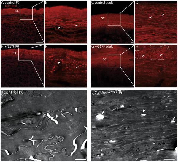
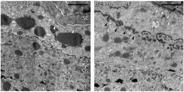
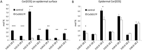
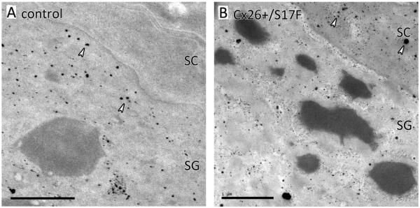
The keratitis-ichthyosis-deafness (KID) syndrome is caused by mutations in the gap junctional channel protein connexin 26 (Cx26), among them the mutation Cx26S17F. Heterozygous Cx26S17F mice resemble the human KID syndrome, i.e. exhibiting epidermal hyperplasia and hearing impairments. Newborn Cx26S17F mice show a defective epidermal water barrier as well as altered epidermal lipid secretion and location. Linoleoyl ω-esterified ceramides are strongly decreased on the skin surface of Cx26S17F mice. Moreover, the epidermal calcium gradient is altered in the mutant mice. These alterations may be caused by an abnormal Cx26S17F channel function that leads to a defective epidermal water barrier, which in turn may trigger the hyperproliferation seen in the KID syndrome.

Keywords: Connexin 26; Epidermal calcium gradient; Epidermal ceramides; Epidermal water barrier defect; Keratitis–ichthyosis–deafness syndrome; Transgenic mouse mutant.


Figures

Fig. 1
Fig. 1
Lipid distribution in the epidermis. (A–D) Nile red stainings of paw epidermis of newborn (A, B) and adult (C, D) control mice. Examples of the evenly distributed lipid lamellae of the stratum corneum are indicated by arrows in B and D. (E–H) In newborn (E, F) and adult (G, H) Cx26S17F mice the lipids accumulate in a dot-like pattern in the stratum corneum (arrows). The stratum corneum of adult mice is strongly thickened leading to fields of “lipid-dots” (surrounded by a dashed line in H). SC: stratum corneum. Scale bars: 20 μm. (I–J) Electron micrographs of stratum corneum of paws. (I) The stratum corneum of control mice shows lipid lamellae in between the corneocytes (arrows). (J) In Cx26S17F mice the stratum corneum appears more compact than control stratum corneum. There are lipid inclusions inside the corneocytes (arrows). Additionally, aggregates of non-processed keratohyalin can be observed (asterisk). Scale bars: 1 μm.
Fig. 2
Fig. 2
Epidermal lipid secretion. (A) Fusion of the lamellar bodies of control mice takes place at the SG/SC junction (white arrowheads). (B) In Cx26S17F mice an additional premature lipid secretion was observed in the SG (black arrowheads). SC: stratum corneum; SG: stratum granulosum. Scale bars: 2 μm.
Fig. 3
Fig. 3
Mass spectrometric analyses of Cer[EOS]. (A) Cer[EOS] with fatty acid residues ranging from h30:0-18:2 to h36:0-18:2 on epidermal surface. The relative amount of Cer[EOS] (in mol% of all ceramides) is strongly diminished on the epidermal surface of Cx26S17F mice. n = 3. (B) Cer[EOS] composition (mol% of all ceramides) in lipid extracts from whole epidermis. Relative amounts of Cer[EOS] are only slightly decreased when extracted from all epidermal layers. n = 4. Cer: ceramide; E: esterified with linoleic acid; O: ω-hydroxylated fatty acid; S: sphingosine long chain base.
Fig. 4
Fig. 4
Ioncapture cytochemistry of epidermal calcium. (A) In control mice SG keratinocytes contains many calcium precipitates (arrows), while the extracellular SG/SC junction as well as the SC corneocyte are completely free of calcium. (B) In the Cx26S17F mutant large amounts of calcium are retained in the SC as well as in the extracellular SG/SC junction (arrows). SG: stratum granulosum; SC: stratum corneum; scale bar: 1 μm.

References

    1. Richard G. Connexin disorders of the skin. Clin. Dermatol. 2005;23:23–32. - PubMed
    1. Natsuga K. Epidermal barriers. Cold Spring Harb. Perspect. Med. 2014;4 - PMC - PubMed
    1. Downing DT. Lipid and protein structures in the permeability barrier of mammalian epidermis. J. Lipid Res. 1992;33:301–313. - PubMed
    1. Rabionet M, Gorgas K, Sandhoff R. Ceramide synthesis in the epidermis. Biochim. Biophys. Acta – Mol. Cell Biol. Lipids. 2014;1841:422–434. - PubMed
    1. Bikle DD, Xie Z, Tu C-L. Calcium regulation of keratinocyte differentiation. Expert Rev. Endocrinol. Metab. 2012;7:461–472. - PMC - PubMed

Publication types

Supplementary concepts