. 2015 Jun 16;42(6):1159-70.
doi: 10.1016/j.immuni.2015.05.012. Epub 2015 Jun 9.

OX40 Ligand Contributes to Human Lupus Pathogenesis by Promoting T Follicular Helper Response

Affiliations

OX40 Ligand Contributes to Human Lupus Pathogenesis by Promoting T Follicular Helper Response

Clément Jacquemin et al. Immunity. .

Abstract

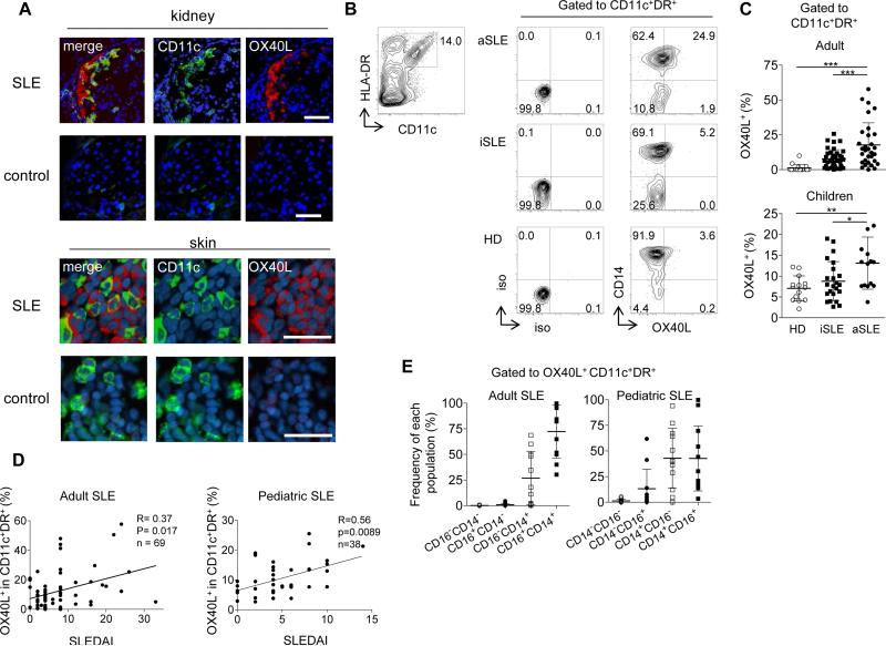
Increased activity of T follicular helper (Tfh) cells plays a major pathogenic role in systemic lupus erythematosus (SLE). However, the mechanisms that cause aberrant Tfh cell responses in SLE remain elusive. Here we showed the OX40 ligand (OX40L)-OX40 axis contributes to the aberrant Tfh response in SLE. OX40L was expressed by myeloid antigen-presenting cells (APCs), but not B cells, in blood and in inflamed tissues in adult and pediatric SLE patients. The frequency of circulating OX40L-expressing myeloid APCs positively correlated with disease activity and the frequency of ICOS(+) blood Tfh cells in SLE. OX40 signals promoted naive and memory CD4(+) T cells to express multiple Tfh cell molecules and were sufficient to induce them to become functional B cell helpers. Immune complexes containing RNA induced OX40L expression on myeloid APCs via TLR7 activation. Our study provides a rationale to target the OX40L-OX40 axis as a therapeutic modality for SLE.


Figures

Fig. 1
Fig. 1. Increased OX40L expression by myeloid APCs in inflammatory tonsils
(A) Expression of OX40L, ICOSL, 4-1BBL and GITRL on myeloid CD11c+HLA-DR+ APCs from pediatric tonsils. A representative result out of 9 independent experiments. (B) Frequency of OX40L+, ICOSL+, GITRL+, and 4-1BBL+ cells within tonsillar myeloid APCs. Mean ± s.d., n=9. One way ANOVA. *** p<0.001. (C) OX40L+CD11c+ APCs in inflammatory tonsils. GC: germinal center; MZ: mantle zone; Epi: Epithelial layers. The scale bars on the top and the bottom panels shows 100 μm and 10 μm, respectively.
Fig. 2
Fig. 2. OX40L expression by myeloid APCs from SLE patients
(A) OX40L+ myeloid APCs in skin and kidney biopsies from adult SLE patients and subjects without autoimmune diseases. A representative result of 5 skin and 3 kidney biopsy samples from SLE patients and 5 skin and 2 kidney biopsy samples from controls. Scale bar=100 μm. (B) Representative flow data on OX40L expression by blood myeloid CD11c+HLA-DR+ APCs from the three groups: healthy donors (HD), inactive (iSLE) and active (aSLE) SLE patients. (C) Frequency of OX40L+ cells within blood myeloid APCs in the three groups in adult and pediatric cohorts. Top: the adult cohort; 16 HD, 38 iSLE, and 31 aSLE samples. Bottom: the children cohort; 14 HD, 20 iSLE, and 14 aSLE samples. One-way ANOVA. * p <0.05, ** p <0.01, *** p <0.001. (D) Correlation between the percentage of OX40L+ cells within CD11c+HLA-DR+ myeloid APCs (adults: n=69 and children: n=38) and disease activity assessed by the SLEDAI. Statistical analysis was performed with the Spearman test. (E) Composition of blood OX40L+ myeloid APCs by different subsets (CD14+CD16-, CD14+CD16+, CD14-CD16-, CD14-CD16+) in adult (n=28) and pediatric (n=34) SLE patients. Mean ± s.d.
Fig. 3
Fig. 3. OX40 signals induce upregulation of Tfh genes
(A) Tfh gene expression by naïve and memory Th cells (from three donors) activated with anti-CD3 and anti-CD28 in the presence or absence of sOX40L for 48 h. Transcript counts in the cultured Th cells are shown after normalization to housekeeping genes. Mean ± s.d., n=3. Paired t-test. * p <0.05, ** p <0.01. (B) Tfh gene expression profiles by naïve and memory Th cells activated with anti-CD3 and anti-CD28 in the presence of indicated reagents for 48 h. Transcript counts in Th cells cultured in the presence of the indicated reagents were normalized to those in control Th cells in each donor. (C) Transcript counts in memory Th cells activated with anti-CD3 and anti-CD28 in the presence of indicated reagents. Mean ± s.d., n=3. One-way ANOVA. * p <0.05, ** p <0.01, *** p <0.001.
Fig. 4
Fig. 4. OX40L stimulation promotes the differentiation of naïve and memory T cells into Tfh-like cells
(A) Expression of CXCR5, IL-21, CD40L, Bcl-6, and T-bet by naïve and memory Th cells activated with anti-CD3 and anti-CD28 in the presence or absence of sOX40L and/or IL-12. Gated to FSChiSSChi activated cells. A representative result out of 3 independent experiments is shown. (B) Frequency of CXCR5+IL-21+,CXCR5+CD40L+, CXCR5+Bcl-6+,and CXCR5+T-bet+ cells developed in naïve or memory Th cells after activation with anti-CD3 and anti-CD28 in the presence or absence of sOX40L and/or IL-12. One-way ANOVA. * p<0.05, ** p<0.01, *** p<0.001, n=3. (C) Naïve or memory Th cells were activated for 4 days with anti-CD3 and anti-CD28 in the presence of sOX40L and/or IL-12, and then cultured with autologous memory B cells. IgG concentrations in the supernatant of each well are shown. A representative result out of 2 independent experiments is shown.
Fig. 5
Fig. 5. Strong TCR stimulation induce naïve Th cells to express Tfh molecules
(A) Bcl-6 expression by naïve Th cells transfected with the indicated siRNA and cultured for 3 days with anti-CD3 and CD28 ± sOX40L. Gated to FSChiSSChi activated cells. Mean ± s.e.m., n=3. (B) Expression of the indicated markers by naïve Th cells activated for 4 days with the indicated number of anti-CD3 and anti-CD28-coated beads. Gated to FSChiSSChi activated cells. A representative result out of 3 independent experiments is shown.
Fig. 6
Fig. 6. The frequency of OX40L+ myeloid APCs correlates with the frequency of ICOS+ blood Tfh cells in human SLE
(A) Expression of ICOS on blood Tfh cells in the three groups; aSLE, iSLE, and HD. A representative flow result is shown. (B) Correlation between the frequency of OX40L+ cells within blood myeloid APCs and the frequency of ICOS+ cells within blood Tfh cells in SLE patients. Spearman correlation test, n=19.
Fig. 7
Fig. 7. RNP-anti-RNP ICs promote OX40L expression by myeloid APCs in a TLR7-dependent manner
(A) Expression of OX40L (MFI) by purified healthy donor monocytes exposed to control sera (n=7) or SLE sera (n=21). Mann-Whitney U-test. ** p <0.01. A representative staining is shown on the left panel. (B) OX40L expression upon stimulation of purified healthy donor monocytes by TLR3, TLR7 or TLR9 agonists. A representative staining out of 4 different experiments is shown. (C) Fold change in OX40L expression (MFI) in healthy donor monocytes exposed to SLE sera (n=7) in the presence or not of a TLR7 inhibitor. Paired t-test. **** p <0.0001. A representative staining is shown on the left panel. (D) OX40L expression (MFI) in healthy donor monocytes exposed to anti-RNPneg SLE sera (n=5) or anti-RNPpos SLE sera (n=16). Mann-Whitney U-test. ** p <0.01. (E) OX40L expression of purified healthy donor monocytes exposed to anti-RNPneg SLE serum, the serum supplemented with anti-RNP-containing IgG, the serum spiked with anti-RNP-containing IgG in the presence of a TLR7 inhibitor. A representative staining out of three independent experiments is shown.

References

    1. Aicher A, Hayden-Ledbetter M, Brady WA, Pezzutto A, Richter G, Magaletti D, Buckwalter S, Ledbetter JA, Clark EA. Characterization of human inducible costimulator ligand expression and function. J Immunol. 2000;164:4689–4696. - PubMed
    1. Akiba H, Takeda K, Kojima Y, Usui Y, Harada N, Yamazaki T, Ma J, Tezuka K, Yagita H, Okumura K. The role of ICOS in the CXCR5+ follicular B helper T cell maintenance in vivo. J Immunol. 2005;175:2340–2348. - PubMed
    1. Barrat FJ, Coffman RL. Development of TLR inhibitors for the treatment of autoimmune diseases. Immunological reviews. 2008;223:271–283. - PubMed
    1. Barrat FJ, Meeker T, Gregorio J, Chan JH, Uematsu S, Akira S, Chang B, Duramad O, Coffman RL. Nucleic acids of mammalian origin can act as endogenous ligands for Toll-like receptors and may promote systemic lupus erythematosus. The Journal of experimental medicine. 2005;202:1131–1139. - PMC - PubMed
    1. Bentebibel SE, Lopez S, Obermoser G, Schmitt N, Mueller C, Harrod C, Flano E, Mejias A, Albrecht RA, Blankenship D, et al. Induction of ICOS+CXCR3+CXCR5+ TH cells correlates with antibody responses to influenza vaccination. Science Translational Medicine. 2013;5:176ra132. - PMC - PubMed

Publication types

MeSH terms