Review
. 2015 Sep;75(1):68-78.
doi: 10.1016/j.cyto.2015.05.014. Epub 2015 Jun 9.

IL-4 and IL-13 signaling in allergic airway disease

Affiliations
Review

IL-4 and IL-13 signaling in allergic airway disease

Naina Gour et al. Cytokine. 2015 Sep.

Abstract

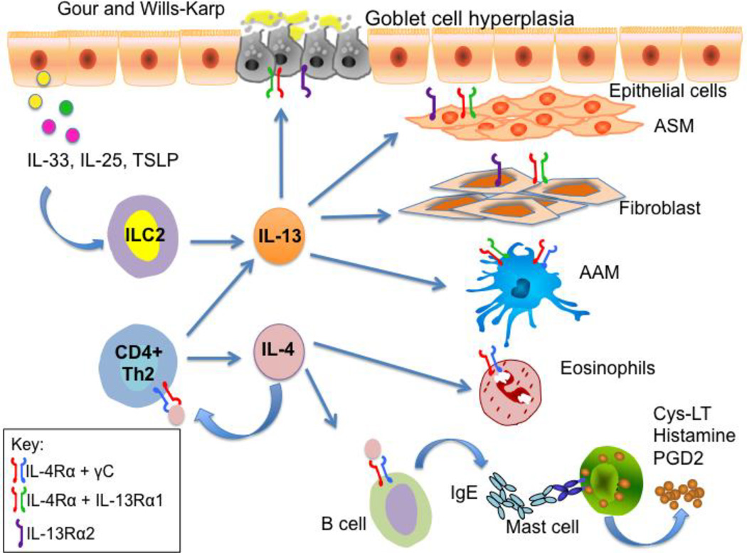
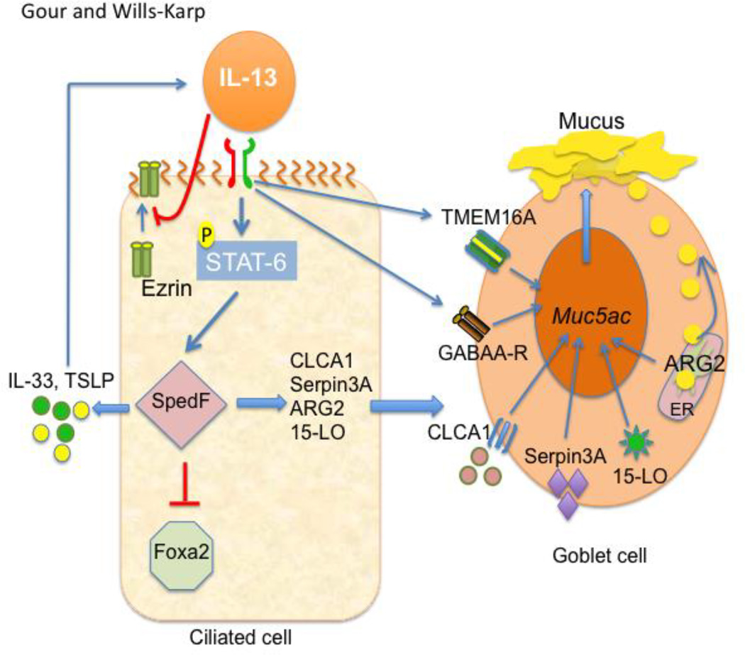
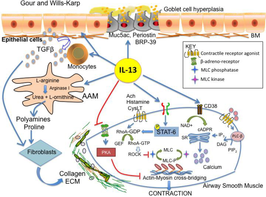
Aberrant production of the prototypical type 2 cytokines, interleukin (IL)-4 and IL-13 has long been associated with the pathogenesis of allergic disorders. Despite tremendous scientific inquiry, the similarities in their structure, and receptor usage have made it difficult to ascertain the distinct role that these two look-alike cytokines play in the onset and perpetuation of allergic inflammation. However, recent discoveries of differences in receptor distribution, utilization/assembly and affinity between IL-4 and IL-13, along with the discovery of unique innate lymphoid 2 cells (ILC2) which preferentially produce IL-13, not IL-4, are beginning to shed light on these mysteries. The purpose of this chapter is to review our current understanding of the distinct roles that IL-4 and IL-13 play in allergic inflammatory states and the utility of their modulation as potential therapeutic strategies for the treatment of allergic disorders.

Keywords: Allergy; Asthma; IL-13; IL-4.


Figures

Figure 1
Figure 1. Differential Roles of IL-4 and IL-13 in Allergic Inflammation
The type 2 cytokines, IL-4 and IL-13 are both produced by CD4+ T cells, whereas only IL-13 is produced by innate lymphoid cells (ILC2s). Through ligation of the type I IL-4R (IL-14Rα/γc) on T cells and mouse B cells, IL-4 preferentially regulates Th2 cell function and IgE synthesis. Although both IL-4 and IL-13 can activate the type II IL-4R (IL-4Rα/IL-13Ra1) on a variety of cells including epithelial cells, airway smooth muscle cells, and fibroblasts, IL-13, not IL-4, mediates the main physiological features of asthma including mucus hypersecretion, subepithelial fibrosis, and airway hyperresponsiveness. IL-13-induced subepithelial fibrosis has been postulated to be mediated via activation of the high affinity IL-13 receptor, IL-13Rα2 on fibroblasts. Cells such as alternatively activated macrophages express both the type I and type II IL-4 receptors.
Figure 2
Figure 2. Potential Mechanisms of IL-13-induced Mucus Cell Metaplasia
Overzealous mucus production is a hallmark of asthma. In the allergic lung, mucus production is predominantly an IL-13-mediated process. IL-13 has been shown to play a pivotal role in several processes important in mucus hypersecretion including: mucus cell metaplasia, mucin gene induction, mucus packaging, and mucus release. The first step is believed to involve the transition of Clara (club) cells to a goblet cell phenotype through the coordinate actions of several transcription factors including Spdef, and Foxa2. IL-13 induces the expression of the Sam pointed domain-containing ETS transcription factor (Spdef) through Type II receptor activation of STAT6-dependent. Spdef, in turn, inhibits the transcription factor, Foxa2 that is required for maintenance of normal differentiation of the airway epithelium. Inhibition of Foxa2 allows induction of a number of genes important in mucus cell metaplasia including: CLCA1, Serpin3A, and 15-lipoxygenase. These genes are thought to directly induce MUC5AC gene expression. On the other hand, anterior gradient 2, Arg2, is thought to play a direct role in mucin folding or another function necessary for maintaining normal mucin production and packaging in the mucus cell endoplasmic reticulum. Other IL-13-inducible genes that play an important role in mucus hypersecretion include: TMEM16A, GABAA-R, and ezrin. Transmembrane protein 16A (TMEM16A), which is a calcium-activated chloride channel, that plays a role in muc5ac expression and mucus release. The γ-aminobutyric acid receptor (GABAAR), which is a pentameric chloride channel, is also thought to regulate mucin gene induction and mucus secretion. Lastly, IL-13 decreases ciliary beat frequency by interfering with the apical localization of the actin binding protein ezrin. This cycle of events is further perpetuated by the ability of Spdef to induce the production of IL-13-promoting cytokines such as IL-33, and TSLP in the epithelium.
Figure 3
Figure 3. Potential Mechanisms of IL-13-induced Airway Hyperresponsiveness
Interleukin 13 induces several cellular changes in the airway wall that may contribute to the main physiological manifestation of asthma, airway hyperresponsiveness. These include: 1) mucus cell metaplasia and mucus production; 2) airway smooth muscle contraction; and 3) collagen deposition and fibrosis. Through STAT6-mediated processes, IL-13 induces a number of epithelial genes including MUC5AC and periostin, which have been shown to result in mucus plugging of the airways and airway hyperresponsiveness. IL-13 also enhances airway smooth muscle sensitivity to a number of contractile agonists (acetylcholine (Ach), histamine, and leukotrienes) through a series of alterations in ASM signaling including: increasing sensitivity to contractile agonist stimulation through enhancing RhoA-ROCK activation and increased myosin light chain phosphorylation, increases in CD38-mediated calcium release, which culminate in actin-myosin cross-bridging and contraction. Lastly, IL-13-induces collagen deposition, through inducing the production of arginase I in alternatively activated macrophages (AAM) and inducing epithelial cell and monocyte production of TGF-β, which induces collagen production from fibroblasts leading to stiffening of the airway wall. RhoA, small GTP binding protein A; ROCK, Rho-associated kinase; GEF, RhoA guanine nucleotide exchange factor.

References

    1. Mosmann TR, et al. Two types of murine helper T cell clone. I. Definition according to profiles of lymphokine activities and secreted proteins. J Immunol. 1986;136(7):2348–2357. - PubMed
    1. Wills-Karp M. Immunologic basis of antigen-induced airway hyperresponsiveness. Annu Rev Immunol. 1999;17:255–281. - PubMed
    1. Wills-Karp M. Interleukin-13 in asthma pathogenesis. Immunol Rev. 2004;202:175–190. - PubMed
    1. Coyle AJ, et al. Interleukin-4 is required for the induction of lung Th2 mucosal immunity. Am J Respir Cell Mol Biol. 1995;13(1):54–59. - PubMed
    1. Brusselle G, et al. Allergen-induced airway inflammation and bronchial responsiveness in wild-type and interleukin-4-deficient mice. Am J Respir Cell Mol Biol. 1995;12(3):254–259. - PubMed

Publication types