. 2015 Aug;138(Pt 8):2173-90.
doi: 10.1093/brain/awv153. Epub 2015 Jun 11.

Recessive nephrocerebellar syndrome on the Galloway-Mowat syndrome spectrum is caused by homozygous protein-truncating mutations of WDR73

Affiliations

Recessive nephrocerebellar syndrome on the Galloway-Mowat syndrome spectrum is caused by homozygous protein-truncating mutations of WDR73

Robert N Jinks et al. Brain. 2015 Aug.

Abstract

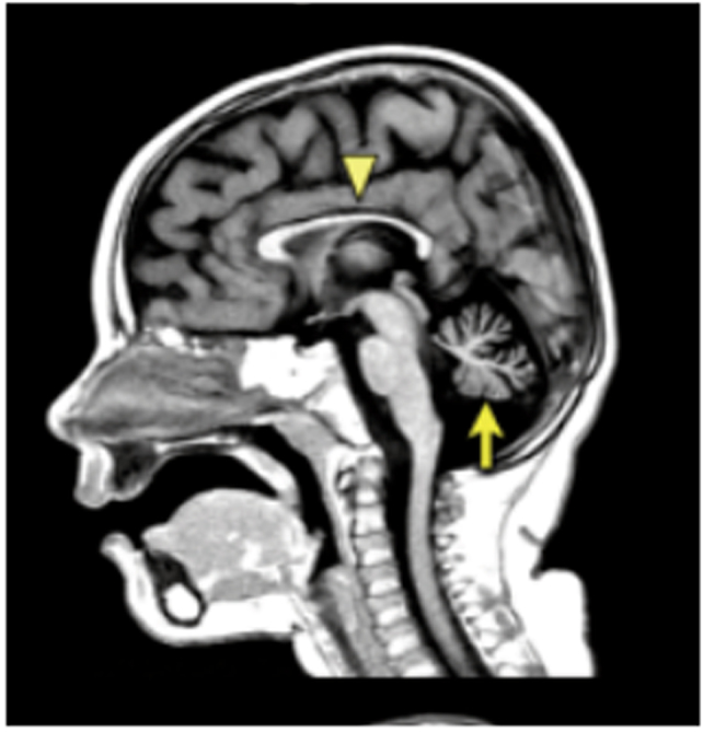
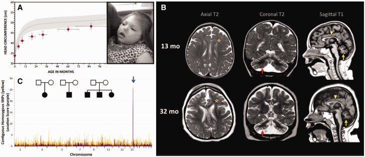
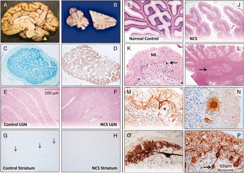
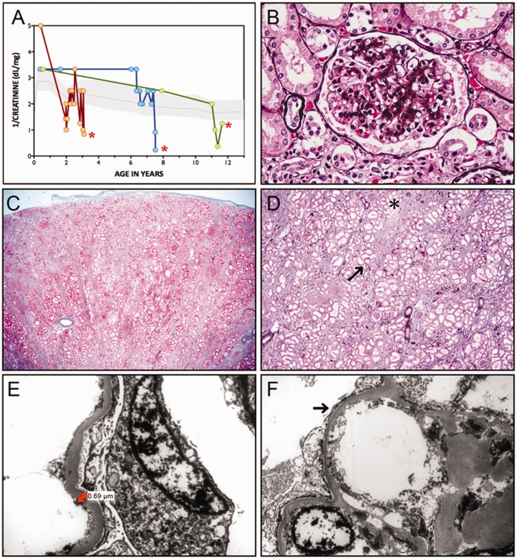
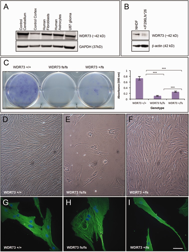
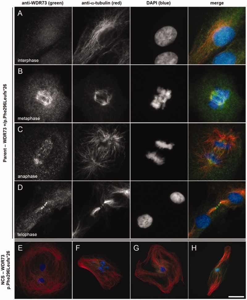
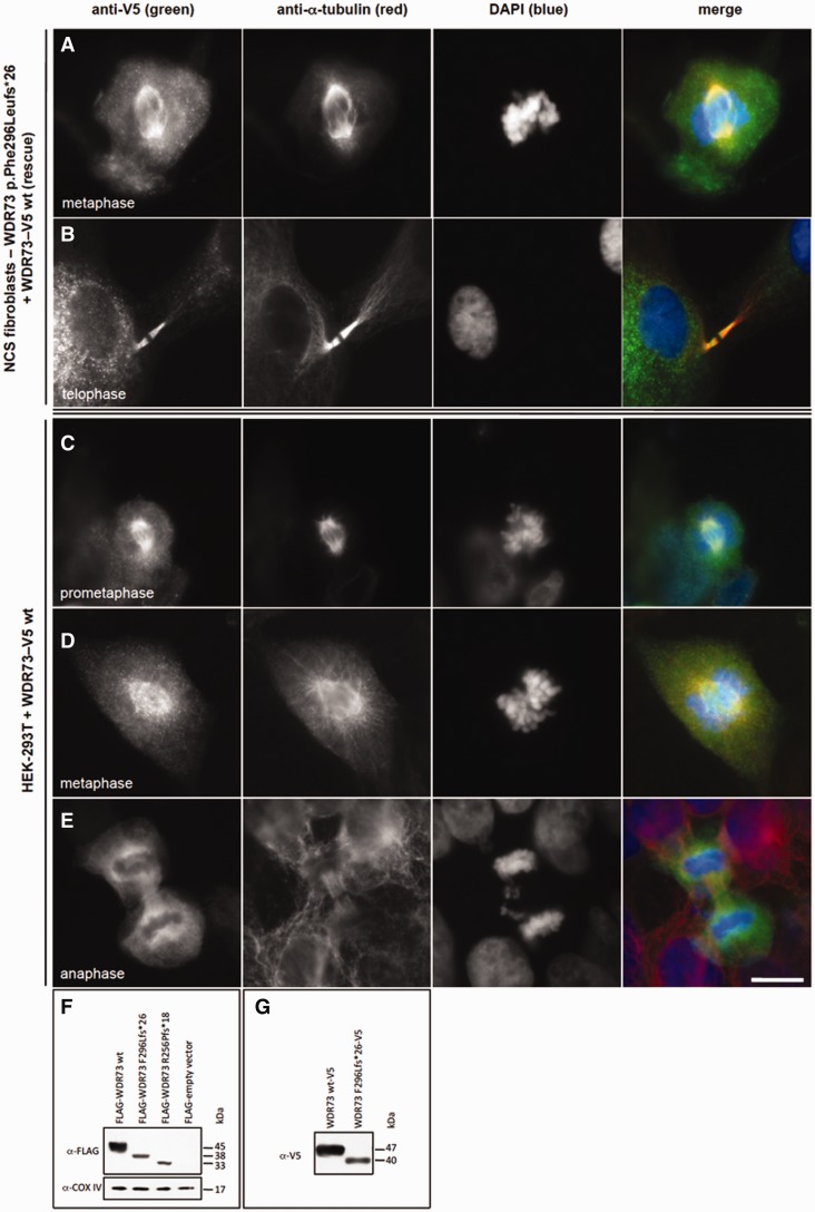
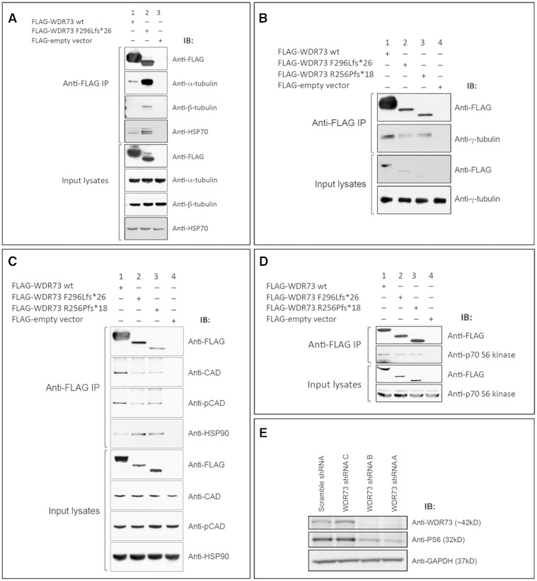
We describe a novel nephrocerebellar syndrome on the Galloway-Mowat syndrome spectrum among 30 children (ages 1.0 to 28 years) from diverse Amish demes. Children with nephrocerebellar syndrome had progressive microcephaly, visual impairment, stagnant psychomotor development, abnormal extrapyramidal movements and nephrosis. Fourteen died between ages 2.7 and 28 years, typically from renal failure. Post-mortem studies revealed (i) micrencephaly without polymicrogyria or heterotopia; (ii) atrophic cerebellar hemispheres with stunted folia, profound granule cell depletion, Bergmann gliosis, and signs of Purkinje cell deafferentation; (iii) selective striatal cholinergic interneuron loss; and (iv) optic atrophy with delamination of the lateral geniculate nuclei. Renal tissue showed focal and segmental glomerulosclerosis and extensive effacement and microvillus transformation of podocyte foot processes. Nephrocerebellar syndrome mapped to 700 kb on chromosome 15, which contained a single novel homozygous frameshift variant (WDR73 c.888delT; p.Phe296Leufs*26). WDR73 protein is expressed in human cerebral cortex, hippocampus, and cultured embryonic kidney cells. It is concentrated at mitotic microtubules and interacts with α-, β-, and γ-tubulin, heat shock proteins 70 and 90 (HSP-70; HSP-90), and the carbamoyl phosphate synthetase 2/aspartate transcarbamylase/dihydroorotase multi-enzyme complex. Recombinant WDR73 p.Phe296Leufs*26 and p.Arg256Profs*18 proteins are truncated, unstable, and show increased interaction with α- and β-tubulin and HSP-70/HSP-90. Fibroblasts from patients homozygous for WDR73 p.Phe296Leufs*26 proliferate poorly in primary culture and senesce early. Our data suggest that in humans, WDR73 interacts with mitotic microtubules to regulate cell cycle progression, proliferation and survival in brain and kidney. We extend the Galloway-Mowat syndrome spectrum with the first description of diencephalic and striatal neuropathology.

Keywords: cerebellar hypoplasia; mTOR; mitosis; nephrosis; progressive microcephaly.


Figures

None
Galloway-Mowat syndrome (GMS) is a neurodevelopmental disorder characterized by microcephaly, cerebellar hypoplasia, nephrosis, and profound intellectual disability. Jinks et al. extend the GMS spectrum by identifying a novel nephrocerebellar syndrome with selective striatal cholinergic interneuron loss and complete lateral geniculate nucleus delamination, caused by a frameshift mutation in WDR73.
Figure 1
Figure 1
Nephrocerebellar syndrome. (A) Postnatal brain growth is slow in children with NCS (grey-shaded area = normal head circumference for age, mean ± 2 SD; red circles = NCS head circumference, mean ± SD). Inset: Children with NCS have no distinctive dysmorphic features, but the forehead characteristically recedes, reflecting relative hypoplasia of the cerebral cortex; post-mortem brain weights are only 50–60% of normal (photos used with parental permission). (B) MRI at 1.5 T (left to right: axial T2, coronal T2, sagittal T1) shows underdeveloped frontal lobes (asterisks), thin corpus callosum (yellow arrowheads), and severe atrophy of the cerebellar hemispheres (red arrows) and vermis (yellow arrows). Comparable images of the same NCS child, taken at 13 months (upper panel) and 32 months (lower panel), reveal progressive degeneration of cerebellar tissue (arrows). (C) Initial SNP genotyping of five children (inset) mapped recessive NCS to an 8.26 Mb autozygous block of DNA on chromosome 15 (blue arrow). After the initial mapping, we identified nine additional patients that allowed us to narrow the shared homozygous interval to 3.96 Mb. Yellow signal indicates the number of contiguous SNPs shared among NCS individuals. For each block of shared homozygous SNPs, purple signal shows the cumulative two-point LOD score (‘location score’), a relative probability, based on population allele frequencies, that the disease gene resides in the homozygous block. Subsequent genotyping of all 27 NCS individuals refined the shared locus to 700 kb, which contained the novel WDR73 c.888delT mutation homozygous in all affected children.
Figure 2
Figure 2
Neuropathology of NCS. (A) A child with NCS died from complications of renal failure at 3.5 years of age. Total brain weight was 636 g (60% of expected) and the hindbrain weighed 7.9% of the total (expected 12%). (B) The cerebellum (yellow arrowhead in A) was small, firm and sclerotic. Luxol Fast Blue (C) and GFAP (D) stains show atrophy and gliosis within cross-sections of the optic nerve. (E) The normal hexalaminar structure of the lateral geniculate nucleus (LGN) is compared with the lateral geniculate nucleus of an NCS child (F), that is nearly devoid of magnocellular neurons, parvocellular neurons, and laminae. (G) Choline acetyltransferase staining of normal striatum reveals several large cholinergic interneurons (arrows), which are absent from a comparable histological section of NCS striatum (H). (I) Normal cerebellar cortex stained with haematoxylin-eosin is compared to that of NCS cerebellum (J), which has short, stubby folia with sparse nuclei in the granule cell layer. (K) Higher magnification shows severe depletion of granule cells with relative preservation of Purkinje neurons (arrow) and a thin, hypercellular molecular layer (ML). (L) Dentate nuclei (arrow) have normal structure and cellularity. (M) Deafferented Purkinje neurons (asterisk) stained for calbindin and neurofilament have ‘weeping’ dendrite configurations, profusion of dendritic elements into ‘asteroid bodies’ (N) and other complex branching patterns (O), and (P) bulbous (‘torpedo’) swelling within proximal axons (arrow).
Figure 3
Figure 3
Kidney disease. (A) Variable onset of nephrosis is followed by progressive loss of glomerular function, shown as 1/serum creatinine (dl/mg), in three with NCS who died (red asterisks) from complications of renal failure at ages 3.5, 8, and 12 years. (B) Jones’ stain reveals both global and segmental sclerosis of glomeruli. (C) ‘Striped’ fibrosis is seen throughout the kidney interstitium. (D) At higher magnification, linear tracks of fibrosis (arrow) correspond to adjacent tubular atrophy (asterisk). (E) Electron micrographs show a 3- to 4-fold thickening of the glomerular basement membrane (normal for age, 0.15 µm) accompanied by (F) effacement and microvillus transformation of podocyte foot processes (arrow).
Figure 4
Figure 4
WDR73 expression in human brain and NCS cells. (A) Immunoblot of WDR73 expression in: lanes 1 and 2, normal post-mortem human cerebellum, cerebral cortex; lanes 3–5, cultured human cells as indicated. (B) Immunoblot of WDR73 from NHDFs and dermal fibroblasts from a heterozygous (WDR73 +/p.Phe296Leufs*26) parent of a child with NCS. Note the additional lower molecular mass band for the truncated WDR73 in the heterozygous parent’s cell lysate. (C) NCS (p.Phe296Leufs*26) fibroblasts (fs/fs) grow poorly in primary culture. Left: Crystal violet cell proliferation assay demonstrating reduced proliferation of dermal fibroblasts from a child with NCS (WDR73 fs/fs) relative to the heterozygous parent’s cells (WDR73 +/fs) and NHDF (WDR73 +/+) plated at the same density. Right: Quantification of cell proliferation by absorbance of extracted crystal violet at 590 nm (n = 4) (***P < 0.001). (D–F) Phase contrast comparison of growth characteristics and morphologies of NHDF (WDR73 +/+), NCS (p.Phe296Leufs*26; WDR73 fs/fs), and heterozygous parent fibroblasts (p.Phe296Leufs*26; WDR73 +/fs) seeded at equal densities and grown under the same conditions on the same 6-well plate. (G–I) Anti-β-actin immunofluorescence microscopy of NHDF (WDR73 +/+), NCS (p.Phe296Leufs*26; WDR73 fs/fs), and heterozygous parent fibroblasts (p.Phe296Leufs*26; WDR73 +/fs). Nuclei were counterstained with DAPI. Scale bars: D–F = 140 µm; G–I = 55 µm.
Figure 5
Figure 5
WDR73 immunoreactivity in fibroblasts from a child with NCS and a heterozygous parent. (A–D) Heterozygous (WDR73 +/p.Phe296Leufs*26) parent fibroblasts. (A) Anti-WDR73 (green) immunoreactivity is diffuse and cytosolic during interphase (anti-α-tubulin, red). (B) WDR73 immunoreactivity localizes to the spindle poles and spindle microtubules during metaphase. (C) WDR73 immunoreactivity at the mitotic spindle poles, kinetochore microtubules and central spindle microtubules during anaphase. (D) WDR73 immunoreactivity localizes to the midbody microtubules apposing the Flemming body (stem body) during telophase. (E–H) WDR73 immunoreactivity is weak and cytosolic in NCS fibroblasts homozygous for WDR73 p.Phe296Leufs*26. (E and F) Twenty-two per cent of NCS fibroblasts observed were binucleate and an additional 20% displayed abnormal nuclear morphology (bi-/multi-lobed nuclei, budding micronuclei, or nucleoplasmic bridges). (G and H) Additional NCS fibroblasts displaying the range of microtubule network morphology observed. Note that the magnifications are reduced in E–H. Scale bars: A–D = 10 µm; E–F = 50 µm; G–H = 90 µm.
Figure 6
Figure 6
Recombinant WDR73-V5 fusion protein rescues cell cycle defect in NCS patient fibroblasts. (A and B) Anti-V5 immunofluorescence (green) demonstrates that WDR73 C-terminal V5 fusion protein (WDR73–V5) overexpressed in NCS patient fibroblasts localizes to the mitotic microtubules (anti-α-tubulin) during metaphase (A) and telophase (B), rescuing the cell cycle defect in these cells. (C–E) Overexpression of WDR73 C-terminal V5 fusion protein (WDR73-V5) in HEK-293T cells. Anti-V5 immunofluorescence is in green; anti-α-tubulin in red. During pro-metaphase (C) and metaphase (D) recombinant WDR73-V5 colocalizes with α-tubulin at the mitotic spindle and aster microtubules. (E) WDR73-V5 localizes to the spindle poles, the kinetochore microtubules and the midzone microtubules during anaphase. Scale bars: A–E = 10 µm. (F–G) Western blots of recombinant N-terminal FLAG WDR73 fusion proteins (F) and WDR73 C-terminal V5 fusion proteins (G) overexpressed for 40–48 h in HEK-293T cells. Anti-COX IV was labelled as a protein loading control. The abundance of FLAG-WDR73 p.Phe296Leufs*26 (F296Lfs*26) was 2.6- to 5.6-fold lower and FLAG-WDR73 p.Arg256Profs*18 (R256Pfs*18) was 1.6- to 6.5-fold lower than that of FLAG-WDR73 wild-type (wt) across four replicates despite transfection of equivalent amounts of plasmid DNA, suggesting instability of the truncated proteins.
Figure 7
Figure 7
WDR73 interacts with α-, β-, and γ-tubulin, CAD, HSP-70, HSP-90, and p70 S6 kinase. (A) FLAG-WDR73 F296Lfs*26 (38 kDa) overexpressed in HEK-293T cells co-immunoprecipitated (IP) substantially more endogenous α- and β-tubulin and HSP-70 than FLAG-WDR73 wild-type (45 kDa). (B) FLAG-WDR73 wild-type, F296Lfs*26, and R256Pfs*18 each co-immunoprecipitated γ-tubulin at an abundance roughly proportional to abundance of the respective WDR73 construct. (C and D) Co-immunoprecipitation of (C) endogenous CAD, phospho-Ser1859 CAD, HSP-90, and (D) p70 S6 kinase from lysates of HEK-293T cells overexpressing the WDR73 constructs indicated. (E) Lentiviral transduction of shRNA plasmid constructs A and B targeting WDR73 in HEK-293FT cells successfully knocked down WDR73 and produced a concomitant decrease in ribosomal protein S6 phosphorylation. GAPDH was labelled as a protein loading control. This immunoblot was labelled with the same anti-WDR73 antibody (Novus) used for immunoblotting throughout the paper (validating the specificity of the antibody).

References

    1. Abraham H, Tornoczky T, Kosztolanyi G, Seress L. Cell formation in the cortical layers of the developing human cerebellum. Int J Dev Neurosci 2001; 19: 53–18. - PubMed
    1. Agromayor M, Martin-Serrano J. Knowing when to cut and run: mechanisms that control cytokinetic abscission. Trends Cell Biol 2013; 23: 433–41. - PubMed
    1. Aosaki T, Miura M, Suzuki T, Nishimura K, Masuda M. Acetylcholine-dopamine balance hypothesis in the striatum: an update. Geriatr Gerontol Int 2010; 10 (Suppl 1): S148–57. - PubMed
    1. Baez S, Couto B, Herrera E, Bocanegra Y, Trujillo-Orrego N, Madrigal-Zapata L, et al. Tracking the Cognitive, social, and neuroanatomical profile in early neurodegeneration: type III cockayne syndrome. Front Aging Neurosci 2013; 5: 80. - PMC - PubMed
    1. Barisoni L. Podocyte biology in segmental sclerosis and progressive glomerular injury. Adv Chronic Kidney Dis 2012; 19: 76–83. - PubMed

Publication types

MeSH terms

Supplementary concepts