. 2015 Sep:156:68-78.
doi: 10.1016/j.exppara.2015.06.003. Epub 2015 Jun 9.

IL-17A promotes protective IgA responses and expression of other potential effectors against the lumen-dwelling enteric parasite Giardia

Affiliations

IL-17A promotes protective IgA responses and expression of other potential effectors against the lumen-dwelling enteric parasite Giardia

Sara M Dann et al. Exp Parasitol. 2015 Sep.

Abstract

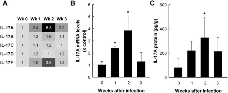
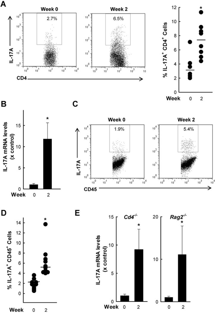
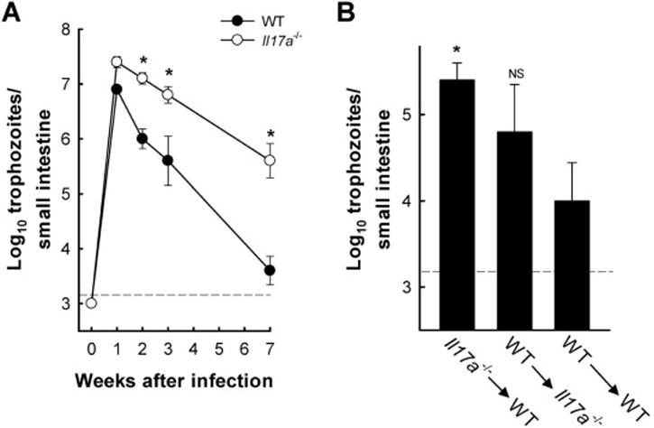
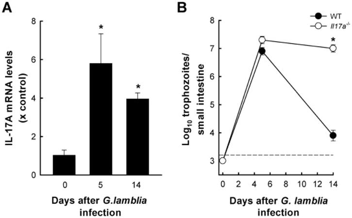
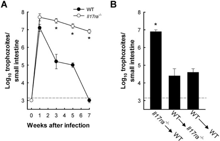
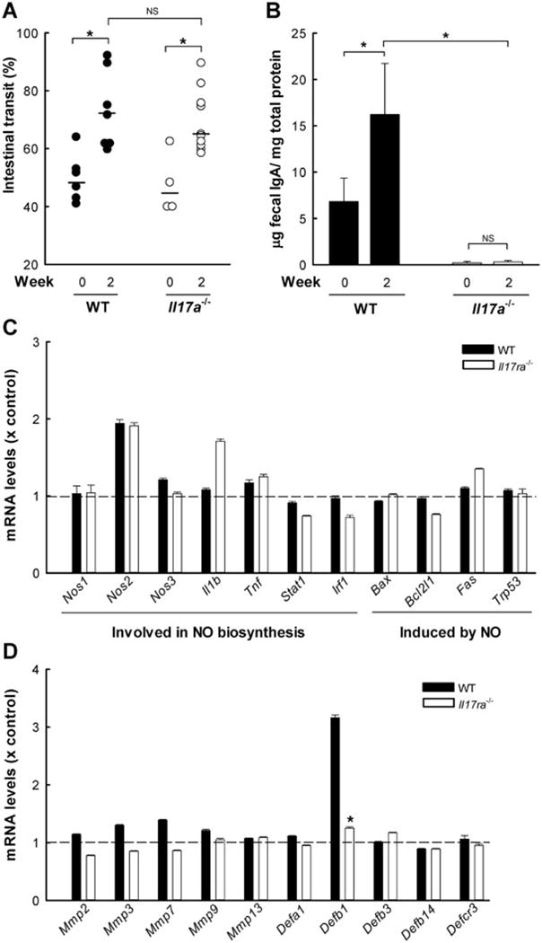
Giardia lamblia is a leading protozoan cause of diarrheal disease worldwide. It colonizes the lumen and epithelial surface of the small intestine, but does not invade the mucosa. Acute infection causes only minimal mucosal inflammation. Effective immune defenses exist, yet their identity and mechanisms remain incompletely understood. Interleukin (IL)-17A has emerged as an important cytokine involved in inflammation and antimicrobial defense against bacterial pathogens at mucosal surfaces. In this study, we demonstrate that IL-17A has a crucial function in host defense against Giardia infection. Using murine infection models with G. muris and G. lamblia, we observed marked and selective induction of intestinal IL-17A with peak expression after 2 weeks. Th17 cells in the lamina propria and innate immune cells in the epithelial compartment of the small intestine were responsible for the IL-17A response. Experiments in gene-targeted mice revealed that the cytokine, and its cognate receptor IL-17RA, were required for eradication of the parasite. The actions of the cytokine were mediated by hematopoietic cells, and were required for the transport of IgA into the intestinal lumen, since IL-17A deficiency led to marked reduction of fecal IgA levels, as well as for increased intestinal expression of several other potential effectors, including β-defensin 1 and resistin-like molecule β. In contrast, intestinal hypermotility, another major antigiardial defense mechanism, was not impacted by IL-17A loss. Taken together, these findings demonstrate that IL-17A and IL-17 receptor signaling are essential for intestinal defense against the important lumen-dwelling intestinal parasite Giardia.

Keywords: Cytokines; Mucosal immunity; Protozoan parasites; Small intestine.


Figures

Figure 1
Figure 1. Increased IL-17A expression after G. muris infection
C57BL/6 mice were orally infected with 104 G. muris cysts, and the small intestine was removed at the indicated times. Uninfected mice were used as controls (Week 0). (A) Heat map of IL-17 family member mRNA expression determined by microarray analysis. Numbers represent the fold increases relative to uninfected controls (Wk 0). Data represent RNA pooled from 6 mice/group. (B) mRNA expression was analyzed by qPCR for IL-17A. Levels are presented as mean + SE of three separate experiments, which each experiment performed on total RNA pooled from 3–5 mice/time point. (C) Levels of IL-17A protein were assessed by ELISA in tissue extracts of the small intestine. Data represent mean + SE of 5–6 mice/time point. Data are representative of results from two independent experiments. *p<0.05 relative to uninfected controls.
Figure 2
Figure 2. Cellular sources of intestinal IL-17A in response to G. muris infection
C57 mice were orally infected with G. muris cysts and examined after two weeks. Uninfected mice were used as a control. (A) CD4+ T cells were isolated from the lamina propria of the small intestine and analyzed by flow cytometry for CD4 and IL-17A expression. Representative FACS dot plots (left) and percentages of IL-17A producing CD4+ cells of individual mice (right). Horizontal bars indicate means. (B–D) Cells were isolated from the epithelial compartment of the small intestine of groups of 3–4 mice, and assessed for IL-17A mRNA expression by qPCR (B; bar graphs represent mean + SE for three independent experiments) and by flow cytometry for co-staining of IL-17A and CD45 (representative FACS dot plot in C, and data on cell percentages from 8–10 individual mice in D; Horizontal bars indicate means). (E) Quantitative PCR analysis of IL-17A mRNA in the small intestine of CD4-deficient (Cd4−/−) and Rag 2-deficient (Rag2−/−) mice infected with G muris. Bar graphs show fold induction (mean + SE; n=3–4) compared to uninfected controls. Data from A–D are representative of the results from three independent experiments; and data from E is representative of the results from two independent experiments; *p <0.05 compared to uninfected controls.
Figure 3
Figure 3. G. muris clearance is impaired in the absence of IL-17A
(A) IL-17A-deficient (open circles) and wild-type (closed circles) mice were infected with G muris, and trophozoite numbers in the small intestine were determined at the indicated times. Data are mean ± SE (n ≥6 mice/time point) from three independent experiments; *p<0.05 compared to wild-type controls. (B) Bone marrow chimeric mice were generated by isolating and transferring bone marrow cells from Il17a−/− mice into C57BL/6 (WT) mice (Il17a−/−→WT), from WT to Il17a−/− mice (WT→Il17a−/−), or as controls from WT to WT mice (WT→WT). Following an 8 week reconstitution period, mice were infected orally with G muris cysts. Trophozoite numbers were determined 7 weeks after infection. Data are mean + SE (n≥6 mice/time point) from two independent experiments; *p<0.05 relative to WT→WT controls. The black dashed lines depict the detection limit of the assays.
Figure 4
Figure 4. IL-17A deficiency impairs eradication of G. lamblia
(A) Wild-type C57BL/6 mice were orally infected with 106 G lamblia GS/M trophozoites. Total RNA was extracted from the small intestine and analyzed for IL-17A mRNA expression by qPCR. Data are mean + SE (n=3–4 mice/time point) from two independent experiments, *p<0.05 relative to uninfected controls (Day 0). (B) Mice deficient in IL-17A (Il17a−/−, open circles) and wild-type (WT, closed circles) mice were infected with G lamblia, and trophozoite numbers were determined at the indicated times. Data are mean ± SE (n=3–4 mice/time point); *p<0.05 relative to wild-type controls. The black dashed line depicts the detection limit of the assay.
Figure 5
Figure 5. IL-17RA signaling in hematopoietic cells is essential for G. muris eradication
(A) Mice deficient in IL-17RA (IL17ra−/−, open circles) and wild-type (WT, closed circles) mice were infected with 104 G muris cysts, and trophozoite numbers in the small intestine were determined at the indicated times. Data are mean ± SE (n=3–4 mice/time point) from three independent experiments; *p<0.05 relative to uninfected controls (Day 0). (B) Bone marrow chimeric mice were generated by isolating and transferring bone marrow cells from IL17ra−/− mice into C57BL/6 (WT) mice (IL17ra−/−→WT), from WT to IL17ra−/− mice (WT→ IL17ra−/−), or as controls from WT to WT mice (WT→WT). Following an 8 week reconstitution period, mice were infected orally with G muris cysts, and trophozoite numbers were determined after 5 weeks. Data are mean + SE (n≥6 mice/time point) from three independent experiments; *p<0.05 relative to WT→WT chimeras. The black dashed lines depict the detection limit of the assays.
Figure 6
Figure 6. Mechanisms of IL-17A signaling-dependent antigiardial defense
IL-17A-deficient (Il17a−/−, open circles and bars in A and B), 17RA-deficient mice (IL17ra−/−, open bars in C and D), and wild-type mice (WT, closed circles and bars in A–D) were infected with 104 G. muris cysts or left uninfected as controls (Week 0), and analyzed two weeks after infection. (A) Small intestinal motility was evaluated by determining the distance traveled by a carmine dye-containing test meal relative to the length of the entire small intestine over a 20-minute period. Each point represents an individual animal from three independent experiments. Horizontal bars indicate medians; *p<0.05 relative to indicated control; NS, not significant compared to wild-type controls. (B) IgA levels were determined in stool homogenates by ELISA and normalized to total protein. Results are mean + SE (n=5–6 mice/time point) from two independent experiments; *p<0.05 relative to uninfected controls. (C, D) Microarray analysis of small intestinal expression of the indicated genes after G. muris infection. Bars show relative expression compared to the respective uninfected control mice. Error bars represent SD for genes with array replicates (n=3–12 replicates/gene); *p<0.05 relative to wild-type controls.

References

    1. Agarwal S, Mayer L. Diagnosis and treatment of gastrointestinal disorders in patients with primary immunodeficiency. Clin Gastroenterol Hepatol. 2013;11:1050–1063. - PMC - PubMed
    1. Aley SB, Zimmerman M, Hetsko M, Selsted ME, Gillin FD. Killing of Giardia lamblia by cryptdins and cationic neutrophil peptides. Infect Immun. 1994;62:5397–5403. - PMC - PubMed
    1. Aley SB, Zimmerman M, Hetsko M, Selsted ME, Gillin FD. Killing of Giardia lamblia by cryptdins and cationic neutrophil peptides. Infect Immun. 1994;62:5397–5403. - PMC - PubMed
    1. Algood HM, Allen SS, Washington MK, Peek RM, Jr, Miller GG, Cover TL. Regulation of gastric B cell recruitment is dependent on IL-17 receptor A signaling in a model of chronic bacterial infection. J Immunol. 2009;183:5837–5846. - PMC - PubMed
    1. Andersen YS, Gillin FD, Eckmann L. Adaptive immunity-dependent intestinal hypermotility contributes to host defense against Giardia spp. Infect Immun. 2006;74:2473–2476. - PMC - PubMed

MeSH terms