. 2015 Jul 15;128(14):2423-9.
doi: 10.1242/jcs.166561. Epub 2015 Jun 12.

Alternative routes to the cell surface underpin insulin-regulated membrane trafficking of GLUT4

Affiliations

Alternative routes to the cell surface underpin insulin-regulated membrane trafficking of GLUT4

Dimitrios Kioumourtzoglou et al. J Cell Sci. .

Abstract

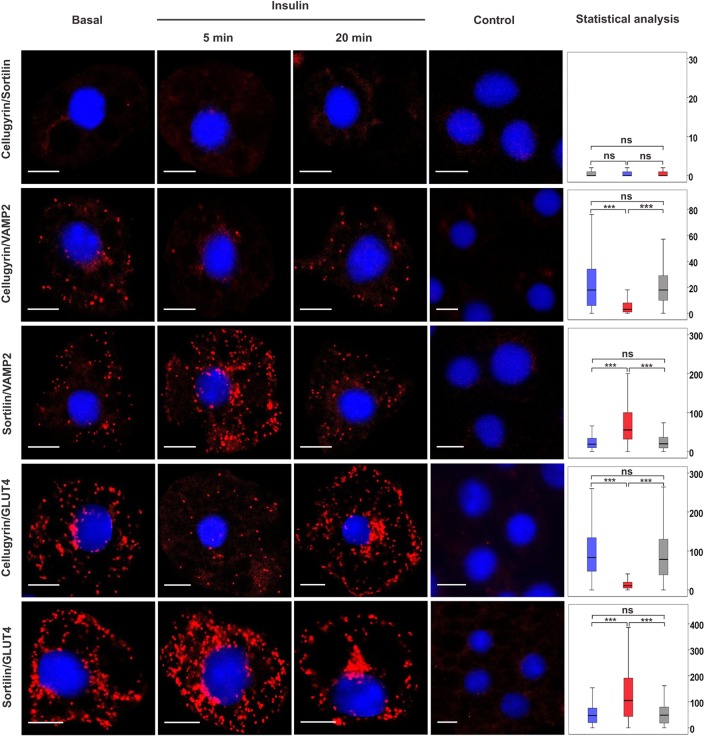
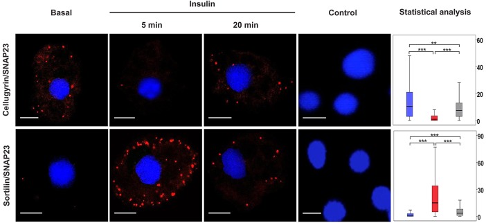
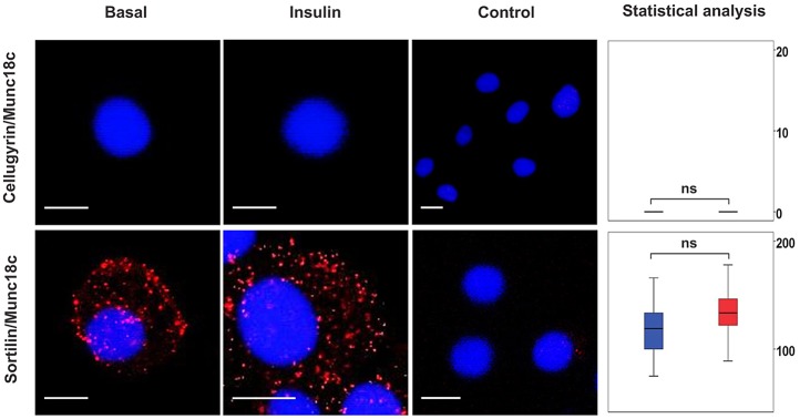
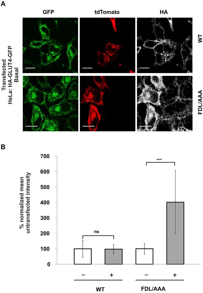
Insulin-stimulated delivery of glucose transporters (GLUT4, also known as SLC2A4) from specialized intracellular GLUT4 storage vesicles (GSVs) to the surface of fat and muscle cells is central to whole-body glucose regulation. This translocation and subsequent internalization of GLUT4 back into intracellular stores transits through numerous small membrane-bound compartments (internal GLUT4-containing vesicles; IGVs) including GSVs, but the function of these different compartments is not clear. Cellugyrin (also known as synaptogyrin-2) and sortilin define distinct populations of IGV; sortilin-positive IGVs represent GSVs, but the function of cellugyrin-containing IGVs is unknown. Here, we demonstrate a role for cellugyrin in intracellular sequestration of GLUT4 in HeLa cells and have used a proximity ligation assay to follow changes in pairwise associations between cellugyrin, sortilin, GLUT4 and membrane trafficking machinery following insulin-stimulation of 3T3-L1 adipoctyes. Our data suggest that insulin stimulates traffic from cellugyrin-containing to sortilin-containing membranes, and that cellugyrin-containing IGVs provide an insulin-sensitive reservoir to replenish GSVs following insulin-stimulated exocytosis of GLUT4. Furthermore, our data support the existence of a pathway from cellugyrin-containing membranes to the surface of 3T3-L1 adipocytes that bypasses GSVs under basal conditions, and that insulin diverts traffic away from this into GSVs.

Keywords: Endosome; Gyrin; Membrane traffic.


Conflict of interest statement

Competing interests

The authors declare no competing or financial interests.

Figures

Fig. 1.
Fig. 1.
Pairwise associations between cellugyrin, sortilin and VAMP2 or GLUT4 in the presence and absence of insulin stimulation. A PLA was used to detect pairwise associations between cellugyrin or sortilin and VAMP2 or GLUT4 in 3T3-L1 adipocytes treated or not (Basal) with 100 nM insulin for 5 or 20 min as indicated (Insulin). PLA signals are shown in red; DAPI stain is in blue. Controls omitting the first listed primary antibody are shown (controls omitting either and both primary antibodies were performed in parallel with no significant signal detected). Statistical analyses of PLA data were performed using Blobfinder and SPSS software. The boxplots show the median number of signals and interquartile range of 200–300 cells per condition (y-axis: blue, basal; red, 5 min insulin stimulation; grey, 20 min insulin stimulation); the upper and the lower whiskers show the 25th and the 75th percentiles, respectively. Images are representative of three independent experiments. For all control experiments, the median PLA signal value was <1 per cell. Any median signal >1 obtained in the presence of both primary antibodies was found to be significantly greater than that obtained in controls for all combinations shown. ns, P≥0.05, ***P<0.001. Scale bars: 10 μm.
Fig. 2.
Fig. 2.
Associations of SNAP23 with cellugyrin and sortilin in the presence and absence of insulin stimulation. A PLA was used to detect pairwise associations of SNAP23 with either cellugyrin or sortilin in 3T3-L1 adipocytes treated or not (Basal) with 100 nM insulin for 5 or 20 min, as indicated (Insulin). PLA signals are shown in red; DAPI stain is in blue. Images are representative of three independent experiments. Data were analysed as for Fig. 1 (y-axis: blue, basal; red, 5 min and grey, 20 min insulin stimulation respectively). ns, P≥0.05, ***P<0.001, **0.001≤P<0.05. Scale bars: 10 μm.
Fig. 3.
Fig. 3.
Pairwise associations between cellugyrin, sortilin and Munc18c in the presence and absence of insulin stimulation. A PLA was used to detect pairwise associations between cellugyrin, sortilin and Munc18c in 3T3-L1 adipocytes treated or not (Basal) with 100 nM insulin for 5 min (Insulin). PLA signals are shown in red; DAPI stain in is blue. Data were analysed and represented as for Fig. 1 (y-axis: blue, basal; red, 5 min and grey, 20 min insulin stimulation respectively). ns, P≥0.05. Scale bars: 10 μm.
Fig. 4.
Fig. 4.
Mutation of a short sorting sequence within the N-terminal cytosolic tail of cellugyrin (FDL to AAA) results in mislocalization to the plasma membrane and loss of intracellular sequestration of GLUT4. A HeLa cell line stably expressing HA–GLUT4–GFP was created following infection with a lentiviral construct encoding GFP-tagged GLUT4 carrying an HA epitope in the first extracellular loop (Muretta et al., 2008). These were subsequently transiently transfected with an expression vector encoding either wild-type (WT) or mutant (FDL/AAA) tdTomato-tagged cellugyrin. (A) Indirect immunofluorescence was used to detect HA–GLUT4–GFP at the plasma membrane by staining in the absence of cell permeabilization (pseudo-coloured, white). Expression and localization of the tdTomato-tagged cellugyrin construct is shown in red and the total amount of HA–GLUT4–GFP in green. Images are representative of three independent experiments. Scale bars: 10 μm. (B) Quantification of indirect HA–GLUT4–GFP immunofluorescence (pseudo-coloured, white) at the plasma membrane was normalized to total GLUT4–GFP fluorescence from cells transfected with either WT or FDL/AAA tdTomato-tagged cellugyrin-encoding vectors (see A). The ratio of surface:total GLUT4 staining is compared between transfected (+) and non-transfected (−) cells from the same coverslip, and shows that surface levels of HA–GLUT4–GFP are increased in cells expressing FDL/AAA cellugyrin. Results represent mean±s.d. from 10 different cells (data were statistically analysed in pairs using a two-tailed Student's t-test; ns, P≥0.05; ***P<0.001.). Quantification of GFP signal from cells expressing either WT or FDL/AAA cellugyrin revealed no difference in total GLUT4–GFP levels between the two (supplementary material Fig. S3).

References

    1. Aran V., Bryant N. J. and Gould G. W. (2011). Tyrosine phosphorylation of Munc18c on residue 521 abrogates binding to Syntaxin 4. BMC Biochem. 12, 19 10.1186/1471-2091-12-19 - DOI - PMC - PubMed
    1. Bai L., Wang Y., Fan J., Chen Y., Ji W., Qu A., Xu P., James D. E. and Xu T. (2007). Dissecting multiple steps of GLUT4 trafficking and identifying the sites of insulin action. Cell Metab. 5, 47-57. 10.1016/j.cmet.2006.11.013 - DOI - PubMed
    1. Belfort G. M., Bakirtzi K. and Kandror K. V. (2005). Cellugyrin induces biogenesis of synaptic-like microvesicles in PC12 cells. J. Biol. Chem. 280, 7262-7272. 10.1074/jbc.M404851200 - DOI - PubMed
    1. Bryant N. J. and Gould G. W. (2011). SNARE proteins underpin insulin-regulated GLUT4 traffic. Traffic 12, 657-664. 10.1111/j.1600-0854.2011.01163.x - DOI - PubMed
    1. Bryant N. J., Govers R. and James D. E. (2002). Regulated transport of the glucose transporter GLUT4. Nat. Rev. Mol. Cell Biol. 3, 267-277. 10.1038/nrm782 - DOI - PubMed

Publication types

MeSH terms