Review
. 2015 Sep:153:107-24.
doi: 10.1016/j.pharmthera.2015.06.006. Epub 2015 Jun 11.

Modulation of the tumor vasculature and oxygenation to improve therapy

Affiliations
Review

Modulation of the tumor vasculature and oxygenation to improve therapy

Dietmar W Siemann et al. Pharmacol Ther. 2015 Sep.

Abstract

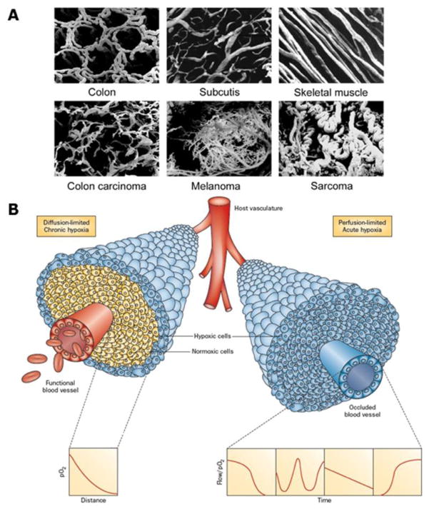
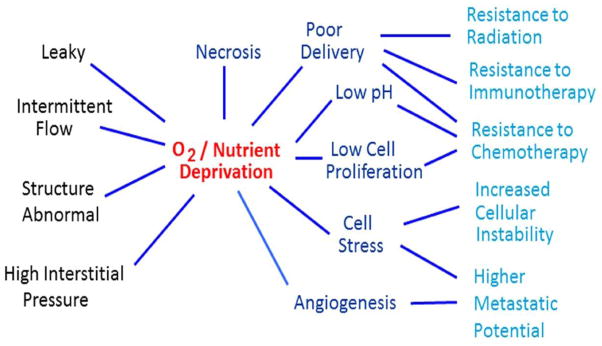
The tumor microenvironment is increasingly recognized as a major factor influencing the success of therapeutic treatments and has become a key focus for cancer research. The progressive growth of a tumor results in an inability of normal tissue blood vessels to oxygenate and provide sufficient nutritional support to tumor cells. As a consequence the expanding neoplastic cell population initiates its own vascular network which is both structurally and functionally abnormal. This aberrant vasculature impacts all aspects of the tumor microenvironment including the cells, extracellular matrix, and extracellular molecules which together are essential for the initiation, progression and spread of tumor cells. The physical conditions that arise are imposing and manifold, and include elevated interstitial pressure, localized extracellular acidity, and regions of oxygen and nutrient deprivation. No less important are the functional consequences experienced by the tumor cells residing in such environments: adaptation to hypoxia, cell quiescence, modulation of transporters and critical signaling molecules, immune escape, and enhanced metastatic potential. Together these factors lead to therapeutic barriers that create a significant hindrance to the control of cancers by conventional anticancer therapies. However, the aberrant nature of the tumor microenvironments also offers unique therapeutic opportunities. Particularly interventions that seek to improve tumor physiology and alleviate tumor hypoxia will selectively impair the neoplastic cell populations residing in these environments. Ultimately, by combining such therapeutic strategies with conventional anticancer treatments it may be possible to bring cancer growth, invasion, and metastasis to a halt.

Keywords: Exercise; Hypoxia modifiers; Metastases; Microenvironment; Stem cells; Vascular targeting.


Conflict of interest statement

Conflict of Interest

The authors declare that there are no conflicts of interest

Figures

Figure 1
Figure 1
A. Vascular casts illustrating the differences in vasculature between normal tissues (colon, subcutis, and skeletal muscle) and malignant tumors (colon, melanoma, and sarcoma). Reprinted with permission from Vaupel. (2004). Tumor microenvironmental physiology and its implications for radiation oncology. Seminars in Radiation Oncology, 14, 198–206. B. Illustration of tumor cells growing as a cord around blood vessels from which they obtain oxygen and nutrients. The left side illustrates oxygen diffusion and utilization from the vessel resulting in the development of chronically hypoxic cells at the outer edge of the cord. The right side shows perfusion through the vessel that has been transiently compromised and results in the development of acute hypoxia; examples of the types of flow/oxygen changes reported during a 60 minute period are illustrated in the 4 panels below. Reprinted with permission from Horsman et al. (2012). Imaging hypoxia to improve radiotherapy outcome. Nature Reviews. Clinical Oncology, 9, 674–687.
Figure 2
Figure 2
A. Micro-regional distribution of hypoxia in SiHa cervix xenograft illustrating co-localization between the bioreductive nitroimidazole agent EF5 (green) and increased expression of the endogenous hypoxia marker HIF-1 (red) at a distance from blood vessels (CD31, blue). Image courtesy of D. Hedley. B. An example of a patient with a FAZA PET positive oropharyngeal cancer Reprinted with permission from Mortensen et al. (2012). FAZA PET/CT hypoxia imaging in patients with squamous cell carcinoma of the head and neck treated with radiotherapy: results from the DAHANCA 24 trial. Radiotherapy and Oncology, 105, 14–20.
Figure 3
Figure 3
The invasive capacity of both TIC and non-TIC sorted from DU145 and PC-3ML human prostate cancer cells increased significantly upon exposure to hypoxia (1% O2, 24 hours). Columns, mean; bars, SD (n=4); **, p<0.01, ***, p<0.001 (t-test).
Figure 4
Figure 4
A. and B. CTSL secreted levels in PC-3ML cells exposed to hypoxic (1% O2) or acidic conditions (pH 6.8) for the indicated durations followed by reoxygenation or restoration of neutral pH conditions for a total time of 24 h. Secreted CTSL levels were determined by ELISA on cell conditioned media and normalized to cell numbers. Shown are mean and standard error values calculated from three independent experiments. *, p<0.05, **, p<0.005, ***, p<0.001. C. and D. Invasive capacities of PC-3ML cells exposed to hypoxia or acidic pH for indicated durations. Mean and standard error are shown. *, p<0.05, **, p<0.005, ***, p<0.001. Modified from Sudhan et al. (2013). Cathepsin L inhibition by the small molecule KGP94 suppresses tumor microenvironment enhanced metastasis associated cell functions of prostate and breast cancer cells. Clinical & Experimental Metastasis, 30, 891–902.
Figure 5
Figure 5
Four clinical trials showing the relationship between tumor oxygenation and outcome of therapy. A. Progression free survival (PFS) for 90 patients with nasopharyngeal tumors treated with chemoradiotherapy and stratified for whether their tumors were hypoxic (high expression of HIF-1α and CA IX) or non-hypoxic. Reprinted with permission from Hui et al. (2002). Coexpression of hypoxia-inducible factors 1alpha and 2alpha, carbonic anhydrase IX, and vascular endothelial growth factor in nasopharyngeal carcinoma and relationship to survival. Clinical Cancer Research, 8, 2595–2604. B. Freedom from biochemical failure for 57 prostate patients treated with brachytherapy in which the prostate/muscle (P/M) mean pO2 ratio estimated using the Eppendorf electrode was above or below 0.10. Reprinted with permission from Turaka et al. (2012). Hypoxic prostate/muscle PO2 ratio predicts for outcome in patients with localized prostate cancer: long-term results. International Journal of Radiation Oncology, Biology, Physics, 82, e433–439. C. Disease-free survival in 40 patients with head & neck squamous cell carcinoma (SCC) based on the pre-radiation therapy estimate of hypoxia as determined by a tumor-to-muscle ratio of > 1.4 from [18F] FAZA PET measurements. Reprinted with permission from Mortensen et al. (2012). FAZA PET/CT hypoxia imaging in patients with squamous cell carcinoma of the head and neck treated with radiotherapy: results from the DAHANCA 24 trial. Radiotherapy and Oncology, 105, 14–20. D. Overall survival in 98 patients with cervical cancer in which the level of perfusion measured with DCE-MRI was either high pre-radiation, low at pre-radiation and subsequently increasing during therapy, or persistently low. Reprinted from Mayr et al. (2010). Longitudinal changes in tumor perfusion pattern during the radiation therapy course and its clinical impact in cervical cancer. International Journal of Radiation Oncology, Biology, Physics, 77, 502–508.
Figure 6
Figure 6
Inherent characteristics of the aberrant tumor blood vessel network result in oxygen and nutrient depravation which negatively impact anticancer therapies and promote tumor cell dissemination resulting in enhanced treatment failures.
Figure 7
Figure 7
Bright field images and corresponding hemoglobin saturation and blood supply time (BST) maps of an untreated Caki-2 renal cell tumor and a tumor receiving daily administration of Sunitinib (oral gavage, 100 mg/kg) over time. Treatment with Sunitinib resulted in an inhibition of tumor vessel development and higher microvessel oxygenation values in comparison to controls. In addition, treatment with Sunitinib resulted in faster BST values in comparison to controls, indicating the formation of a more organized vascular network. Modified from Lee et al. (2014). In vivo spectral and fluorescence microscopy comparison of microvascular function after treatment with OXi4503, Sunitinib and their combination in Caki-2 tumors. Biomedical Optics Express, 5, 1965–1979.
Figure 8
Figure 8
C3H mammary carcinomas in CDF1 mice were locally irradiated with single doses of radiation and the percentage of animals showing tumor control within 90 days were recorded. The radiation dose-response curve, determined from logit analysis, is shown as the dashed line in each figure. Each figure also shows how this radiation response curve is modified by various treatments. A. within 1-hour after irradiating i.p. injecting 50 mg/kg OXi4503 (●) or 20 mg/kg DMXAA (○); B. carbogen breathing for 5 minutes before and during irradiation (●) or an intraperitoneal (i.p.) injection with 1000 mg/kg nicotinamide 30 minutes before irradiating (○); C. i.p. injecting 500 mg/kg nimorazole (○) or doranidazole (●) 30 minutes prior to irradiating; D. irradiating (Rad) tumors in the middle (0h) of heating (HT) at 42.5°C for 60 minutes (●) or irradiating (Rad) tumors and heating (HT) at 42.5°C for 60 minutes 4-hours (4h) later (○). Modified from Horsman et al. (2011). Impact on radiotherapy. In D. W. Siemann (Ed.), Tumor Microenvironment (pp. 353–376). Chichester: John Wiley & Sons, Ltd.
Figure 9
Figure 9
Blood flow measured at rest and during exercise in rat prostate or rat prostate tumor tissue. *p<0.05 tumor in exercised vs resting host; †p<0.05 tumor vs prostate in exercised host. A two-way analysis of variance with repeated measures was used to compare within-group (rest vs exercise) and between-group (control vs tumor-bearing) differences in blood flow. Modified from McCullough et al. (2014). Modulation of blood flow, hypoxia, and vascular function in orthotopic prostate tumors during exercise. Journal of the National Cancer Institute, 106, dju036.
Figure 10
Figure 10
Representative fluorescence photomicrograph of a prostate tumor cross section following injection of EF5 (red; 30 mg/kg) and Hoechst-33342 (blue; 40 mg/kg) to illustrate hypoxia (red) and patent vessels (blue) in sedentary (A) and exercise-trained (B) orthotopic rat prostate tumors. C. and D. are graphic representations of the fraction of tissues bound by EF5 and patent vessel counts respectively. *p<0.05 between groups. Reprinted with permission from McCullough et al. (2013). Effects of exercise training on tumor hypoxia and vascular function in the rodent preclinical orthotopic prostate cancer model. Journal of Applied Physiology, 115, 1846–1854.

References

    1. Adams GE, Cooke MS. Electron-affinic sensitization. I. A structural basis for chemical radiosensitizers in bacteria. International Journal of Radiation Biology and Related Studies in Physics, Chemistry, and Medicine. 1969;15:457–471. - PubMed
    1. Adams GE, Flockhart IR, Smithen CE, Stratford IJ, Wardman P, Watts ME. Electron-affinic sensitization. VII. A correlation between structures, one-electron reduction potentials, and efficiencies of nitroimidazoles as hypoxic cell radiosensitizers. Radiation Research. 1976;67:9–20. - PubMed
    1. Ahn GO, Brown M. Targeting tumors with hypoxia-activated cytotoxins. Frontiers in Bioscience. 2007;12:3483–3501. - PubMed
    1. Alqawi O, Wang HP, Espiritu M, Singh G. Chronic hypoxia promotes an aggressive phenotype in rat prostate cancer cells. Free Radical Research. 2007;41:788–797. - PubMed
    1. Armstrong RB, Laughlin MH. Rat muscle blood flows during high-speed locomotion. Journal of Applied Physiology. 1985;59:1322–1328. - PubMed

Publication types

MeSH terms

Substances