. 2016 Nov;13(6):828-838.
doi: 10.1038/cmi.2015.55. Epub 2015 Jun 15.

Measles virus hemagglutinin triggers intracellular signaling in CD150-expressing dendritic cells and inhibits immune response

Affiliations

Measles virus hemagglutinin triggers intracellular signaling in CD150-expressing dendritic cells and inhibits immune response

Olga Romanets-Korbut et al. Cell Mol Immunol. 2016 Nov.

Erratum in

Abstract

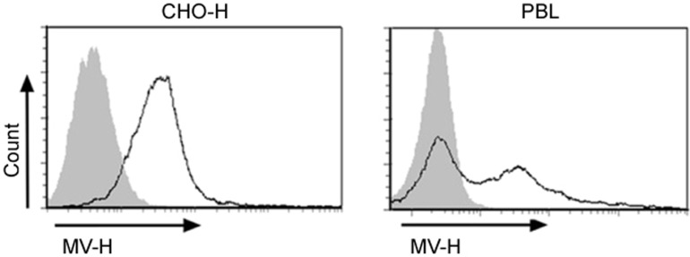
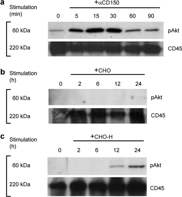
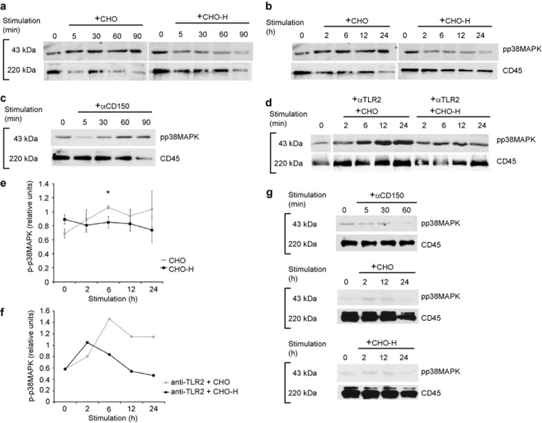
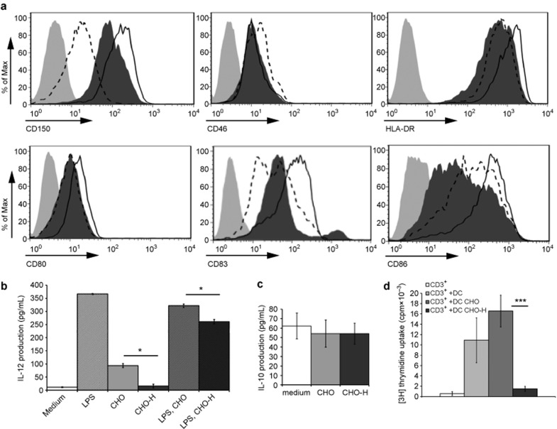
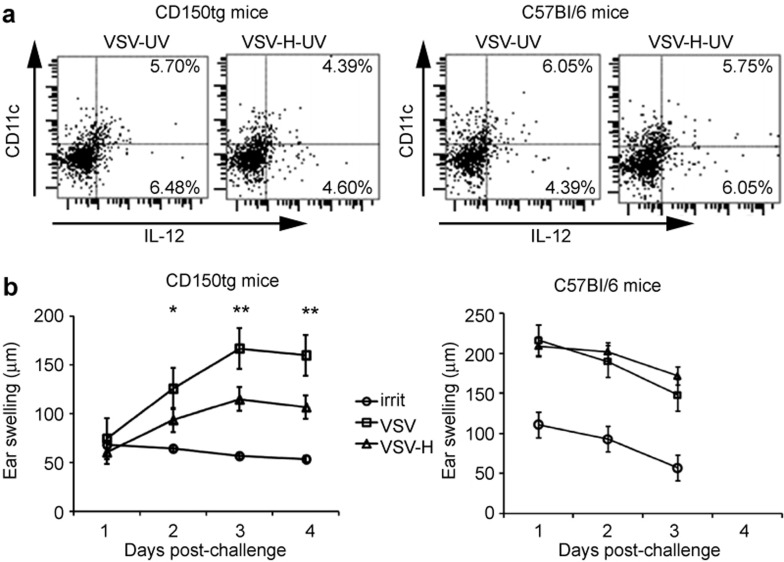
Measles virus (MV) is highly contagious pathogen, which causes a profound immunosuppression, resulting in high infant mortality. This virus infects dendritic cells (DCs) following the binding of MV hemagglutinin (MV-H) to CD150 receptor and alters DC functions by a mechanism that is not completely understood. We have analyzed the effect of MV-H interaction with CD150-expressing DCs on the DC signaling pathways and consequent phenotypic and functional changes in the absence of infectious context. We demonstrated that contact between CD150 on human DCs and MV-H expressed on membrane of transfected CHO cells was sufficient to modulate the activity of two major regulatory pathways of DC differentiation and function: to stimulate Akt and inhibit p38 MAPK phosphorylation, without concomitant ERK1/2 activation. Furthermore, interaction with MV-H decreased the expression level of DC activation markers CD80, CD83, CD86, and HLA-DR and strongly downregulated IL-12 production but did not modulate IL-10 secretion. Moreover, contact with MV-H suppressed DC-mediated T-cell alloproliferation, demonstrating profound alteration of DC maturation and functions. Finally, engagement of CD150 by MV-H in mice transgenic for human CD150 decreased inflammatory responses, showing the immunosuppressive effect of CD150-MV-H interaction in vivo. Altogether, these results uncover novel mechanism of MV-induced immunosuppression, implicating modulation of cell signaling pathways following MV-H interaction with CD150-expressing DCs and reveal anti-inflammatory effects of CD150 stimulation.


Figures

Figure 1
Figure 1
The expression level of MV-H on CHO-H cells and on human PBLs infected by MV. Human PBLs, infected by wt MV (G954 strain) at MOI of 1 for 48 h, and CHO-H transfectants were stained with anti-MV-H mAb cl55 for flow cytometric analysis.
Figure 2
Figure 2
CD150 modulates Akt signaling pathway in human DCs. DCs were co-cultured with CHO/CHO-H cells (a, c–d) at 1:5 ratio or stimulated with 10 μg/mL of anti-CD150 mAbs (clone IPO3) (b) for the different time. Activation of signaling pathways via TLR2 and DC-SIGN receptors was blocked with specific anti-TLR2 (c) and anti-DC-SIGN (d) mAbs. FSL-1 was used as specific ligands for TLR2 stimulation. Cell lysates were analyzed for pAkt (S473) expression by western blot with CD45 as loading control. The level of pAkt was normalized against the level of CD45 using TotalLab program (e, f). (e) The results are expressed as mean (±SD) from 3–6 independent experiments, and differences observed at 6, 12, and 24 h were statistically significant (*P < 0.05, **P < 0.001).
Figure 3
Figure 3
MV-H modulates Akt signaling pathway in human B-lymphoblastoid cell line MP-1 in the absence of an infectious context. MP-1 cells were stimulated by anti-CD150 antibodies IPO3 (a) or co-cultured with CHO (b) or CHO-H (c) cells at 1:5 ratio for the different time. Cell lysates were analyzed for pAkt (S473) expression by western blot with CD45 as loading control. Data represent one of the three independent experiments.
Figure 4
Figure 4
CD150-mediated p38MAPK phosphorylation is decreased upon MV-H-CD150 interaction in DCs. DCs were either co-cultured with CHO/CHO-H cells at 1:5 ratio (a, b) or stimulated with 10 μg/mL of anti-CD150 IPO-3 antibody at different time points (c). Activation of signaling pathways via TLR2 receptor was blocked with specific anti-TLR2 antibodies (d). Cell lysates were analyzed for pp38 MAPK (T180/Y182) expression by western blot with CD45 as a loading control. The level of ppMAPK38 was normalized against the level of CD45 using TotalLab program (e, f). (e) The results are expressed as mean (±SD) from at least three independent experiments and differences observed at 6 h were statistically significant (*P < 0.05, Student's t-test). (g) MP-1 cells were either stimulated with 10 μg/mL of anti-CD150 IPO-3 antibody or co-cultured with CHO/CHO-H cells at 1:5 ratio at different time points.
Figure 5
Figure 5
Interaction of human DCs with MV-H causes changes in DC maturation and function. (a) Downregulation of several surface markers expression in DCs. DCs, co-cultured with CHO (solid line) or CHO-H cells (dashed line) at 1:5 ratio for 24 h, were stained for CD150, CD46, CD80, HLA-DR, CD83, and CD86 (light-gray histogram: nonstained DCs; dark-gray histogram: DCs before CHO/CHO-H co-cultures). Data represent one of the four independent experiments with different donors. IL-12 (b) and IL-10 (c) production by DCs was measured by ELISA in supernatants of either freshly generated DCs (“medium”), or DCs, co-cultured with CHO or CHO-H cells at 1:5 ratio for 24 h (“CHO/CHO-H”). Alternatively, DCs were stimulated by 10 ng/mL LPS for 12 h followed or not by CHO/CHO-H co-culture (“LPS, CHO/CHO-H” or “LPS” respectively). Data represent one of the four independent experiments with different donors. (d) Human peripheral blood CD3-positive T cells (2 × 10⁴) were cultured in quadruplicates for 6 days with culture medium, 104 of UV-irradiated DCs, either freshly generated (DC) or previously co-cultured 24 h with either CHO (DC CHO) or CHO-H (DC CHO-H) at 1:5 ratio. T-cell proliferation was measured on day 6 by [3H] thymidine uptake during last 16 h of culture. Results, expressed as mean cpm (±SD), are from one of 6 representative experiments. The statistical significance was determined by unpaired two-tailed Student's t-test (*P < 0.05, ***P < 0.0001).
Figure 6
Figure 6
CD150 interaction with MV-H inhibits cellular immune response in vivo. (a) Production of IL-12 in DCs isolated from murine lymph nodes after injection with UV-inactivated VSV/VSV-H. Groups of 5 CD150tg and C57B/6 mice were injected with UV-inactivated VSV/VSV-H and 6 h later sensitized with DNFB. DCs were isolated from inguinal, axillary and popliteal lymph nodes 30 h later, double-stained with anti-CD11c and intracellular IL-12 mAb and analyzed on FACS. Data represent one of three independent experiments. (b) Hypersensitivity responses were analyzed in groups of five CD150tg or C57BL/6 mice, injected i.p. with 5 × 10⁶ PFU of VSV-UV or VSV-H-UV. Mice were sensitized 6 h later with DNFB or left unsensitized (irritation “irrit” group) and challenged then 5 days later. Results are expressed as mean ear swelling (±SD) of three experiments at different time points after challenge. A statistical significance was determined by one-way ANOVA (*P < 0.05, **P < 0.001).
Figure 7
Figure 7
Schematic presentation of mechanisms possibly involved in MV-H-induced modulation of the immunobiology of DCs and inhibition of inflammatory immune responses. MV-H interaction with CD150-expressing DCs modifies signaling pathways Akt and p38MAPK (1) and affects DC maturation and function (decreased expression of DC maturation markers, inhibition of IL-12 production and T-cell proliferation) (2). CD150 engagement with MV-H in vivo results in the inhibition of inflammatory responses in humanized CD150tg mice (3). Modulation of cell transduction pathways in DCs and inhibition of their maturation and function are likely to be linked to the suppression of inflammatory responses, seen in measles patients (dashed arrows).

References

    1. Perry RT, Gacic-Dobo M, Dabbagh A et al. Global control and regional elimination of measles, 2000-2012. MMWR Morb Mortal Wkly Rep 2014; 63: 103–107. - PMC - PubMed
    1. Moss WJ, Griffin DE. Measles. Lancet 2012; 379: 153–164. - PubMed
    1. de Swart RL, Ludlow M, de Witte L et al. Predominant infection of CD150+ lymphocytes and dendritic cells during measles virus infection of macaques. PLoS Pathog 2007; 3: e178. - PMC - PubMed
    1. Grosjean I, Caux C, Bella C et al. Measles virus infects human dendritic cells and blocks their allostimulatory properties for CD4+ T cells. J Exp Med 1997; 186: 801–812. - PMC - PubMed
    1. Schnorr JJ, Xanthakos S, Keikavoussi P, Kampgen E, ter Meulen V, Schneider-Schaulies S. Induction of maturation of human blood dendritic cell precursors by measles virus is associated with immunosuppression, Proc Natl Acad Sci U S A 1997; 94: 5326–5331. - PMC - PubMed

Publication types

Substances