. 2015 Jul 7;23(7):1350-61.
doi: 10.1016/j.str.2015.05.006. Epub 2015 Jun 11.

MemProtMD: Automated Insertion of Membrane Protein Structures into Explicit Lipid Membranes

Affiliations

MemProtMD: Automated Insertion of Membrane Protein Structures into Explicit Lipid Membranes

Phillip J Stansfeld et al. Structure. .

Abstract

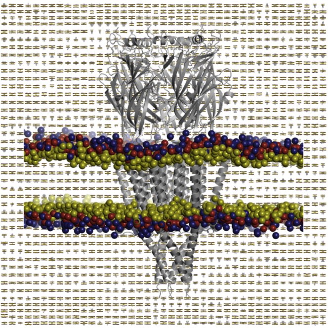
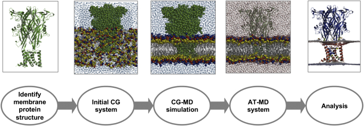
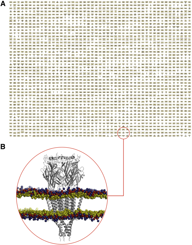
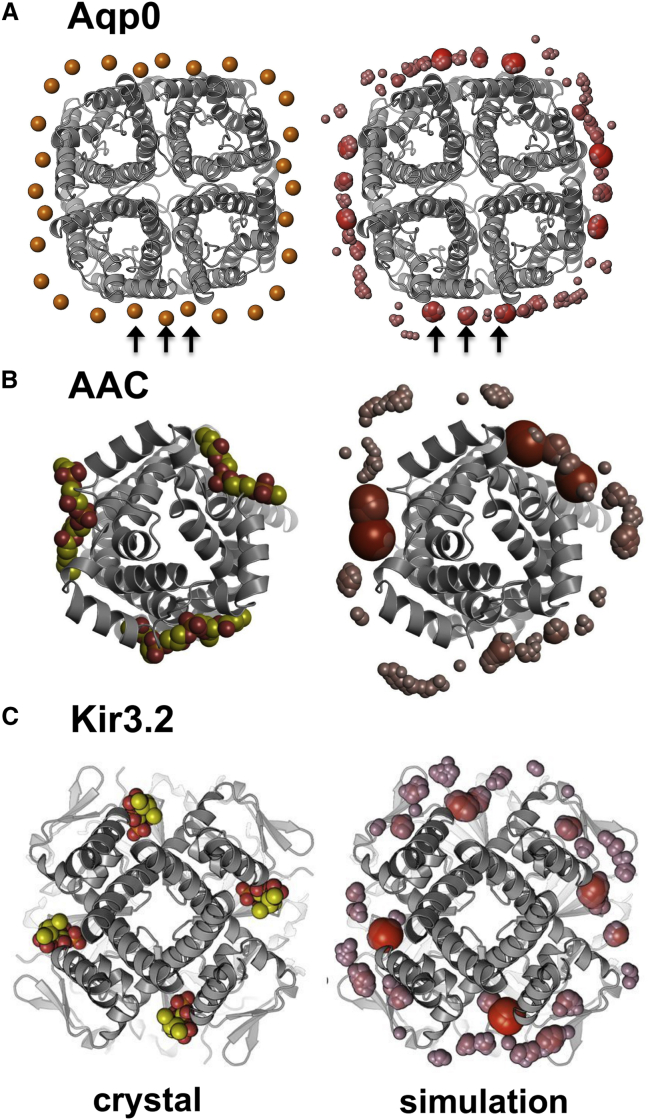
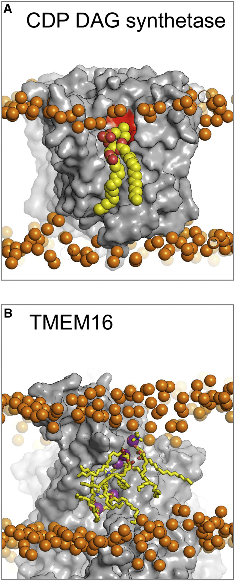
There has been exponential growth in the number of membrane protein structures determined. Nevertheless, these structures are usually resolved in the absence of their lipid environment. Coarse-grained molecular dynamics (CGMD) simulations enable insertion of membrane proteins into explicit models of lipid bilayers. We have automated the CGMD methodology, enabling membrane protein structures to be identified upon their release into the PDB and embedded into a membrane. The simulations are analyzed for protein-lipid interactions, identifying lipid binding sites, and revealing local bilayer deformations plus molecular access pathways within the membrane. The coarse-grained models of membrane protein/bilayer complexes are transformed to atomistic resolution for further analysis and simulation. Using this automated simulation pipeline, we have analyzed a number of recently determined membrane protein structures to predict their locations within a membrane, their lipid/protein interactions, and the functional implications of an enhanced understanding of the local membrane environment of each protein.


Figures

None
Graphical abstract
Figure 1
Figure 1
The MemProtMD Pipeline for Inserting Membrane Proteins into Bilayers The first step is to detect the protein structures from the PDB, here shown for the P2X4 receptor (PDB: 4DW0, trimeric biological assembly as annotated in the PDB). The second is to set up a lipid, water, and protein simulation system. CG simulations are then run (1 μs duration) to assemble and equilibrate a bilayer around each membrane protein structure. The CG simulation system is then converted to atomic resolution, allowing detailed analysis of lipid bilayer/protein interactions. See also Figure S1.
Figure 2
Figure 2
A Database of Membrane Proteins in Phospholipid Bilayers (A) Snapshots of the final (i.e., 1 μs) configurations of 2,294 membrane proteins in a phospholipid (PC) bilayer. (B) Zoomed-in representation of the 5-HT3 ligand-gated ion channel (PDB: 4PIR; pentameric asymmetric unit and biological assembly) in a PC bilayer. Lipid headgroups are shown as spheres representing the choline (blue), phosphate (red), and glycerol (yellow) particles of the CG model. See also Figure S2.
Figure 3
Figure 3
Identifying Lipid Interaction Sites Lipid binding sites for three membrane proteins, identified as regions with a high frequency of occurrence of the phosphate particles of PC molecules. In each case the corresponding crystal structure and MemProtMD simulation snapshot are shown on the left- and right-hand side, respectively. The relative dimensions and redness of the densities reflect the frequency of binding, with those sites that are repeatedly sampled larger in size and redder in color. (A) Aquaporin Aqp0 (PDB: 3M9I) comparing the phosphorus atoms of the headgroups of the experimentally resolved PE lipids (orange spheres) with the phosphate particles from a CG simulation in a PC bilayer. While the phosphate binding sites agree well with the cryo-EM structures (as indicated by the arrows), the phosphates show preference for some sites over others, with a number of the more prominent sites residing at and either side of the subunit interfaces. Note that the biological assembly in the PDB is octameric, so the biological tetramer was regenerated using PISA. See also Figure S5. (B) Cardiolipin molecules bound to the ADP/ATP carrier (AAC; PDB: 4C9G) compared with phosphates from a CG simulation. The level to which the phosphates bind reflects the composition of the binding site and suggests the most favorable interactions with pseudo-subunit X, then Y, then Z. Both yeast and bovine homologs of AAC show differences in the phosphate densities for each binding site. (C) A Kir3.2 channel structure (PDB: 3SYA; tetrameric biological assembly from the PDB) with four bound molecules of a short-tail analog of PIP2, again compared with phosphates from a CG simulation.
Figure 4
Figure 4
Interaction Sites for Lipid-like Ligands (A) The CDP-DAG synthetase (PDB: 4Q2E; dimeric asymmetric unit and biological assembly in the PDB) is involved in a crucial step within the lipid biogenesis pathway. The crystal structures were resolved without bound lipid (gray surface). Here we propose the binding orientation of PC lipids (yellow spheres) to the active site (red surface). While this is not the native substrate or product for this enzyme, the binding reveals the overall orientation of lipid binding to the site. (B) The fungal TMEM16 scramblase (PDB: 4WIS; dimeric asymmetric unit and biological assembly in the PDB) is responsible for permitting lipid flip-flop from one leaflet to the other. MemProtMD is able to predict lipid interaction sites within the permeation pathway, here illustrating five PC lipids (yellow sticks). In this case, not only do we observe lipid binding but also lipid migration, with approximately 15 lipids flipping from one leaflet to the other. Both structures illustrate a snapshot of the lipid binding after 1 μs. Phosphate atoms of the PC lipids are shown as orange spheres.
Figure 5
Figure 5
Local Distortions of the Lipid Bilayer Local distortions of the bilayer induced by membrane proteins may be analyzed by evaluating the positions of the phosphate headgroups of the lipids over the course of the simulations. The extent to which the bilayer deforms can then be compared against the standard DPPC bilayer thickness of 40 Å. The deformations are shown as a mesh on a red-white-blue scale, with points greater than 20 Å from the midplane of the bilayer being colored on a blue gradient and those that are less than 20 Å colored on a red scale. (A) For the enzyme DgkA (PDB: 3ZE5; trimeric asymmetric unit and biological assembly in the PDB), amphipathic helices anchor the protein to one (cytoplasmic) leaflet of the bilayer, such that local distortions are limited to the opposite leaflet. (B) The ABC transporter ABCB10 (PDB: 4AYX; dimeric biological assembly in the PDB) locally perturbs the bilayer such that lipids enter the central core of the protein, suggestive of a proposed pathway for lipid flip-flop catalyzed by related ABC transporters. See also Figure S3.
Figure 6
Figure 6
Amino Acid Distributions Frequencies of occurrence of membrane-interacting amino acid side chains within the TM domains of membrane proteins. The data are shown with respect to the bilayer for all unique α-helical membrane proteins, with the midplane of the bilayer marked as zero and the headgroup region marked by gray lines. In all instances the cytoplasmic side of the membrane (In) relates to the negative distance from the bilayer center of mass. See also Figure S4.
Figure 7
Figure 7
Unexpected Residue Locations (A–D) The observed frequencies of occurrence of residues relative to a bilayer (see Figure 6) may be used as a guideline to identify residues in unusual locations within the membrane. In many instances where residues appear misplaced, bilayer deformations and/or snorkeling of side chains occurs to accommodate charged amino acids within the bilayer (shown as yellow spheres). In some other cases the residue (shown as red spheres) is buried deeply within the bilayer and so is unlikely to be accommodated by such mechanisms. Examples are shown for (A) Lys288 in LeuT (PDB: 4MM8; monomeric biological assembly-1 in the PDB), while Lys196 and Arg453 are able to snorkel; (B) Asn58 in MelB (PDB: 4M64; monomeric biological assembly in the PDB), while Arg52 can escape the hydrophobic core; (C) Lys 972 in SERCA (PDB: 4BEW; monomeric biological assembly in the PDB); and (D) Glu 73 in VDAC1 (PDB: 4C69; monomeric biological assembly in the PDB). In each case the considered protein is monomeric.
Figure 8
Figure 8
Refining Membrane Protein Structures within Membranes Two structures and associated simulations (see main text for details) are shown for the MFS transporter NRT1. (A) A higher-resolution structure (PDB: 4OH3), showing little change in the conformation of TM12 (in blue) on simulation. Both a tyrosine (1) and a tryptophan (2) interact with the headgroups of the lipid bilayer, potentially anchoring the helix. (B) A lower-resolution structure (PDB: 4CL4), showing a (partial) change in register of TM12 (with the red arrows indicating the positions of the helix before and after in the simulation). Only a tyrosine (1) interacts with the membrane in this helix register.

References

    1. Arnarez C., Mazat J.P., Elezgaray J., Marrink S.J., Periole X. Evidence for cardiolipin binding sites on the membrane-exposed surface of the cytochrome bc1. J. Am. Chem. Soc. 2013;135:3112–3120. - PubMed
    1. Badola P., Sanders C.R., 2nd Escherichia coli diacylglycerol kinase is an evolutionarily optimized membrane enzyme and catalyzes direct phosphoryl transfer. J. Biol. Chem. 1997;272:24176–24182. - PubMed
    1. Baeza-Delgado C., Marti-Renom M.A., Mingarro I. Structure-based statistical analysis of transmembrane helices. Eur. Biophys. J. 2013;42:199–207. - PubMed
    1. Bayrhuber M., Meins T., Habeck M., Becker S., Giller K., Villinger S., Vonrhein C., Griesinger C., Zweckstetter M., Zeth K. Structure of the human voltage-dependent anion channel. Proc. Natl. Acad. Sci. USA. 2008;105:15370–15375. - PMC - PubMed
    1. Berman H.M., Westbrook J., Feng Z., Gilliland G., Bhat T.N., Weissig H., Shindyalov I.N., Bourne P.E. The Protein Data Bank. Nucleic Acids Res. 2000;28:235–242. - PMC - PubMed

Publication types

MeSH terms