. 2015;7(5):931-45.
doi: 10.1080/19420862.2015.1055442.

A bi-functional antibody-receptor domain fusion protein simultaneously targeting IGF-IR and VEGF for degradation

Affiliations

A bi-functional antibody-receptor domain fusion protein simultaneously targeting IGF-IR and VEGF for degradation

Yang Shen et al. MAbs. 2015.

Abstract

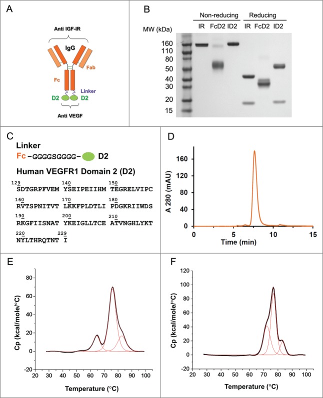
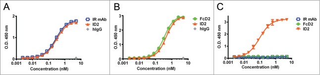
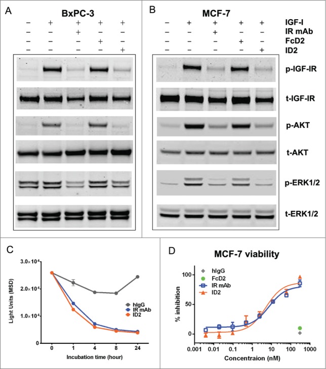
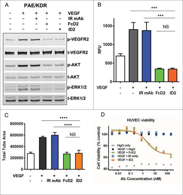
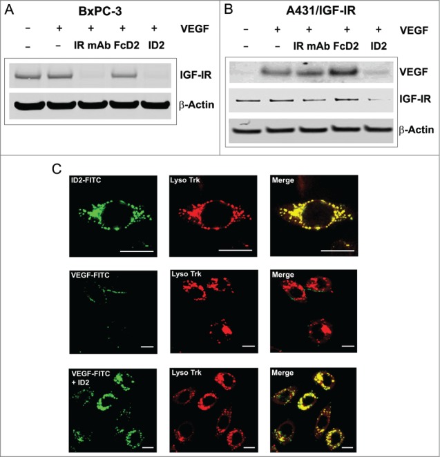
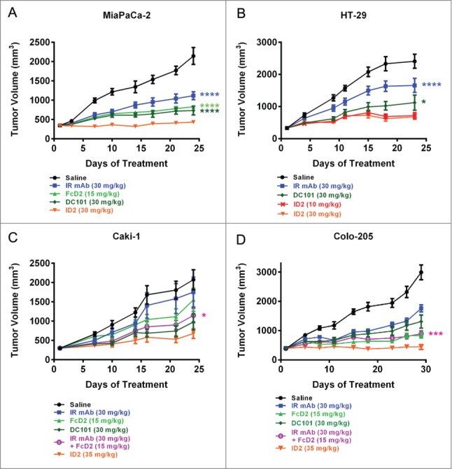
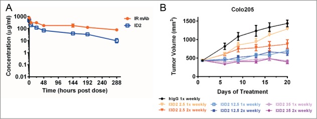
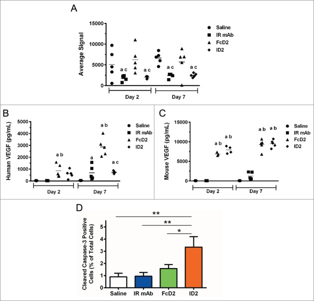
Bi-specific antibodies (BsAbs), which can simultaneously block 2 tumor targets, have emerged as promising therapeutic alternatives to combinations of individual monoclonal antibodies. Here, we describe the engineering and development of a novel, human bi-functional antibody-receptor domain fusion molecule with ligand capture (bi-AbCap) through the fusion of the domain 2 of human vascular endothelial growth factor receptor 1 (VEGFR1) to an antibody directed against insulin-like growth factor - type I receptor (IGF-IR). The bi-AbCap possesses excellent stability and developability, and is the result of minimal engineering. Beyond potent neutralizing activities against IGF-IR and VEGF, the bi-AbCap is capable of cross-linking VEGF to IGF-IR, leading to co-internalization and degradation of both targets by tumor cells. In multiple mouse xenograft tumor models, the bi-AbCap improves anti-tumor activity over individual monotherapies. More importantly, it exhibits superior inhibition of tumor growth, compared with the combination of anti-IGF-IR and anti-VEGF therapies, via powerful blockade of both direct tumor cell growth and tumor angiogenesis. The unique "capture-for-degradation" mechanism of the bi-AbCap is informative for the design of next-generation bi-functional anti-cancer therapies directed against independent signaling pathways. The bi-AbCap design represents an alternative approach to the creation of dual-targeting antibody fusion molecules by taking advantage of natural receptor-ligand interactions.

Keywords: IGF-IR; VEGF; VEGFR1; VEGFR2; angiogenesis; antibody fusion; bi-functional antibody; bispecific antibody; degradation; internalization.


Figures

Figure 1.
Figure 1.
Design and molecular structure of bi-functional antibody receptor domain fusion molecule with VEGF capture (bi-AbCap). (A) Cartoon illustration of the designed bi-AbCap fusion molecule. Orange: Anti IGF-IR IgG backbone, IR mAb; Purple: polypeptide linker; Green: extracellular domain 2 of human VEGFR1 (D2). An FcD2 control molecule was made containing the regions of the hinge, Fc, linker and D2. (B) SDS-PAGE of IR mAb, FcD2 and bi-AbCap ID2. (C) Top: linker sequence: G4SG4; Bottom: the D2 domain used for fusion encompassing amino acids 129–229 of hVEGFR1. (D) ID2 elutes as a mono-disperse peak by size exclusion chromatograpy (SEC). (E) ID2 displays 3 melting temperatures (Tm) when the thermal stability of ID2 measured by differential scanning calorimetry (DSC). Tm1 = 64.4 ± 0.0°C, Tm2 = 76.0 ± 0.0°C, Tm3 = 82.7 ± 0.1°C. (F) IR mAb displays 3 Tms by DSC: Tm1 = 72.2 ± 0.1°C, Tm2 = 76.8 ± 0.0°C, Tm3 = 83.2 ± 0.1°C.
Figure 2.
Figure 2.
Binding to IGF-IR, VEGF and evidence for co-engagement of both targets by ELISA. (A) ID2 (orange), IR mAb (blue) and human IgG control (gray) binding to human IGF-IR-Fc. (B) ID2 (orange), FcD2 (green) and human IgG control (gray) binding to human VEGF. (C) ID2 (orange), IR mAb (blue) and FcD2 (green) binding to both human IGF-IR-Fc (coated) and human VEGF (detected). Each panel is a representative experiment of at least 3 repeated measurements. The graph is plotted as mean ± SEM (n = 2 ).
Figure 3.
Figure 3.
Inhibition of IGF-IR-mediated tumor cell signaling and functions by ID2. (A) 100 nM ID2 inhibits IGF-I-induced phosphorylation of IGF-IR, downstream AKT and ERK1/2 in BxPC-3 cells as assessed by immunoblotting analysis. IR mAb and FcD2 were used as controls. (B) 100 nM of ID2 inhibits IGF-I-induced phosphorylation of IGF-IR, downstream AKT and ERK1/2 in MCF-7 cells in immunoblotting analysis. IR mAb and FcD2 were used as controls. (C) Down regulation of surface IGF-IR on MCF-7 cells when treated with ID2, control IR mAb and control human IgG is measured at 0, 1, 4, 8 and 24 hours by IGF-IR electro-chemiluminescence (ECL) assay. (D) ID2 potently inhibits IGF-I induced MCF-7 viability in a dose dependent manner in a CellTiter Glo assay. The error bar represents the SEM from each triplicate measurement.
Figure 4.
Figure 4.
Inhibition of VEGF-mediated endothelial cell signaling and functions by ID2. (A) 100 nM ID2 inhibits VEGF-induced phosphorylation of VEGFR2, downstream AKT and ERK1/2 in PAE/KDR cells as assessed by immunoblotting analysis. IR mAb and FcD2 were used as controls. (B) In an Oris cell migration assay, PAE/KDR cells stimulated with 100 ng/ml VEGF were treated with 100 nM ID2, IR mAb, or FcD2 for 20 hours. The fluorescence intensity of migrated cells in relative fluorescence units (RFU) was measured. ID2 significantly reduced the migration compared to VEGF and IR mAb controls (p = 0.002 and p = 0.003, respectively, one way ANOVA). (C) ID2 inhibits VEGF stimulated cord formation in an ADSC/ECFC co-culture system. The total tube area for each treatment was calculated. ID2 significantly reduced the total tube area compared with VEGF only and IR mAb controls (p < 0.0001 and p < 0.0001, respectively, one way ANOVA). (D) ID2 inhibits human VEGF induced HUVEC viability in a dose dependent manner in a CellTiter Glo assay. The error bar from panels B, C and D represents the SEM from each triplicate measurement.
Figure 5.
Figure 5.
Evidence for internalization and degradation of both IGF-IR and VEGF by ID2 in vitro. (A) In BxPC-3 cells, ID2 at 20 nM induces IGF-IR internalization and degradation in the presence of 100 ng/mL VEGF by immunoblotting. IR mAb and FcD2 were used as controls. (B) In A431/IGF-IR cells, ID2 at 10 nM induces both surface IGF-IR and supernatant VEGF internalization/degradation in the presence of 400 ng/mL (10 nM calculated as a dimer) VEGF by immunoblotting. IR mAb and FcD2 were used as controls. (C) Fluorescence confocal microscopy analysis on the delivery of ID2 and VEGF to the lysosome in BxPC-3 cells: (top row) the co-localization of FITC-labeled ID2 with lyso tracker; (middle and bottom row) the co-localization of FITC labeled VEGF with the lysosome is dependent on ID2 treatment. Scale bar = 20 μm.
Figure 6.
Figure 6.
ID2 inhibits tumor growth in multiple mouse xenograft models and demonstrates superior anti-tumor activity compared to the IR mAb/FcD2 combination. Tumor bearing mice were treated with controls and ID2 by intraperitoneal injection 3 times per week. The figure shows tumor volume (in mm3) as a function of treatment time (in days). The models tested were: (A) MiaPaCa-2; (B) HT-29; (C) Caki-1; (D) Colo-205. Tumor volume is plotted as mean ± SEM (n = 12). For statistical analysis, RM ANOVA was used through the last day of each study to compare the tumor growth between ID2 and other treatments. Levels of statistical significance are indicated as: *p = 0.01–0.05; ***p = 0.0001–0.001; ****p < 0.0001.
Figure 7.
Figure 7.
In vivo stability of ID2. (A) Pharmacokinetic assessment of serum concentrations of ID2 and IR mAb as a function time (in hours), following 30 mg/kg intraveneous administration in CD-1 mice (n = 3). Total IgG was used for determining the serum concentrations. The serum concentrations were plotted as mean ± SD. (B) Dose frequency study in a Colo-205 mouse xenograft model. 2.5 mg/kg (orange line), 12.5 mg/kg (blue line) and 35 mg/kg (purple line) of I3D2 (an engineered variant of ID2) were dosed intraperitoneally once weekly (light colored line) and twice weekly (dark colored line) schedules. Tumor volumes are plotted as mean ± SEM (n = 12).
Figure 8.
Figure 8.
In vivo mechanism of action in Colo-205 xenograft model. (A) Average signal of total human IGF-IR from lysates of excised tumor, (B) human VEGF concentration (pg/ml) from mouse plasma and (C) Mouse VEGF concentration (pg/ml) from mouse plasma after 2- and 7-day treatments with saline, IR mAb, FcD2 and ID2 were determined by electrochemiluminescent assay. Significant differences in mean (p < 0.05, n = 5) are indicated (a vs Saline; b vs IR mAb; c vs FcD2). (D) Percentage of nuclei with positive immunohistochemistry staining for cleaved caspase-3 from representative tumor tissue after 2-day treatment was compared and plotted as mean ± SEM (n = 5). ID2 treated group had significantly increased cleaved caspase-3 activity compared to saline control (p = 0.0035, one way ANOVA), IR mAb (p = 0.0041, one way ANOVA) and FcD2 (p = 0.0254, one way ANOVA). All charts were generated and statistical analyses were performed with SigmaPlot or Graphpad Prism 6.

References

    1. Chames P, Baty D. Bispecific antibodies for cancer therapy: the light at the end of the tunnel? mAbs 2009; 1:539-47; PMID:20073127; http://dx.doi.org/10.4161/mabs.1.6.10015 - DOI - PMC - PubMed
    1. Kontermann RE, Brinkmann U. Bispecific antibodies. Drug Dis Today 2015; PMID:25728220 - PubMed
    1. Linke R, Klein A, Seimetz D. Catumaxomab: clinical development and future directions. mAbs 2010; 2:129-36; PMID:20190561; http://dx.doi.org/10.4161/mabs.2.2.11221 - DOI - PMC - PubMed
    1. Dong J, Demarest SJ, Sereno A, Tamraz S, Langley E, Doern A, Snipas T, Perron K, Joseph I, Glaser SM, et al.. Combination of two insulin-like growth factor-I receptor inhibitory antibodies targeting distinct epitopes leads to an enhanced antitumor response. Mol Cancer Ther 2010; 9:2593-604; PMID:20716637; http://dx.doi.org/10.1158/1535-7163.MCT-09-1018 - DOI - PubMed
    1. Lu D, Zhang H, Ludwig D, Persaud A, Jimenez X, Burtrum D, Balderes P, Liu M, Bohlen P, Witte L, et al.. Simultaneous blockade of both the epidermal growth factor receptor and the insulin-like growth factor receptor signaling pathways in cancer cells with a fully human recombinant bispecific antibody. J Biol Chem 2004; 279:2856-65; PMID:14576153; http://dx.doi.org/10.1074/jbc.M310132200 - DOI - PubMed

Publication types

MeSH terms