. 2015 Jun 18;161(7):1527-38.
doi: 10.1016/j.cell.2015.05.025. Epub 2015 Jun 11.

ER Stress Sensor XBP1 Controls Anti-tumor Immunity by Disrupting Dendritic Cell Homeostasis

Affiliations

ER Stress Sensor XBP1 Controls Anti-tumor Immunity by Disrupting Dendritic Cell Homeostasis

Juan R Cubillos-Ruiz et al. Cell. .

Abstract

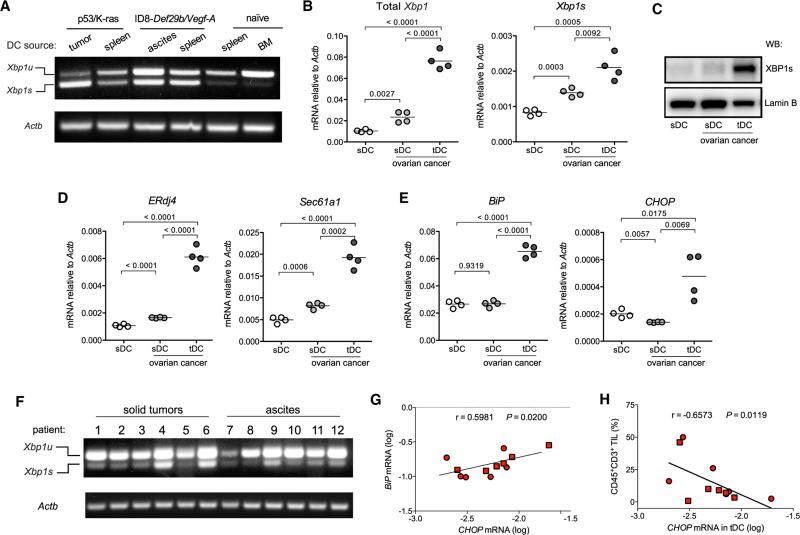
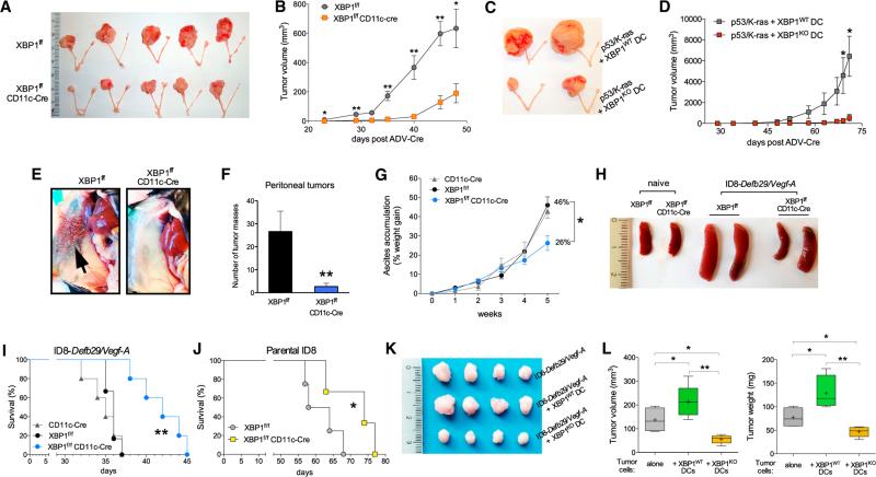
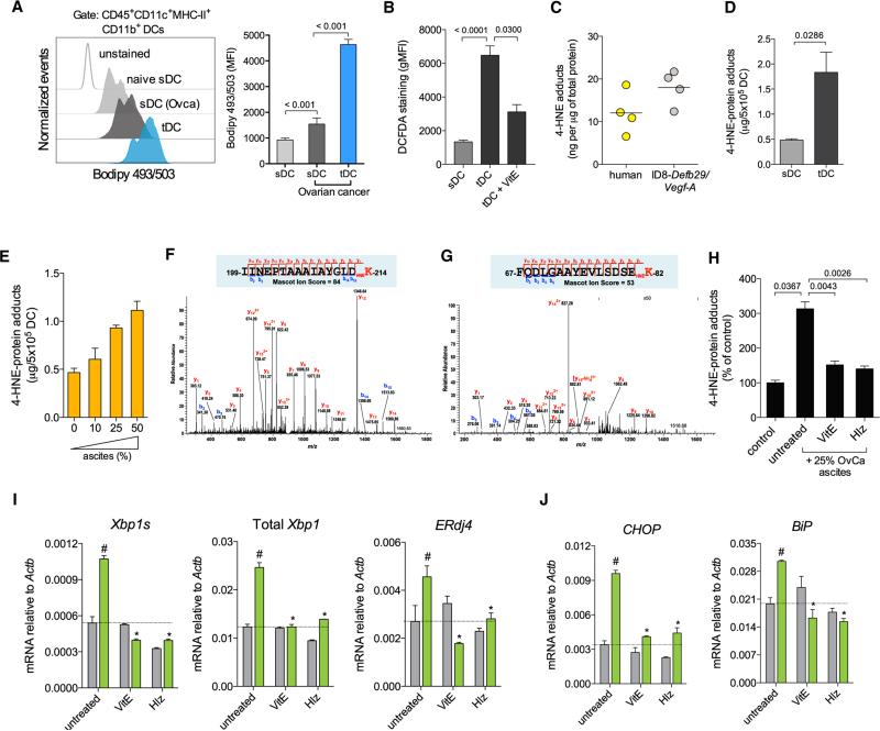
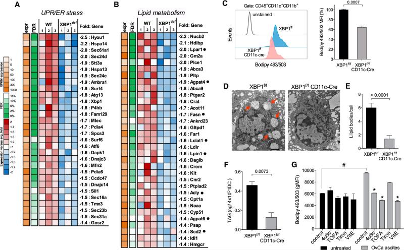
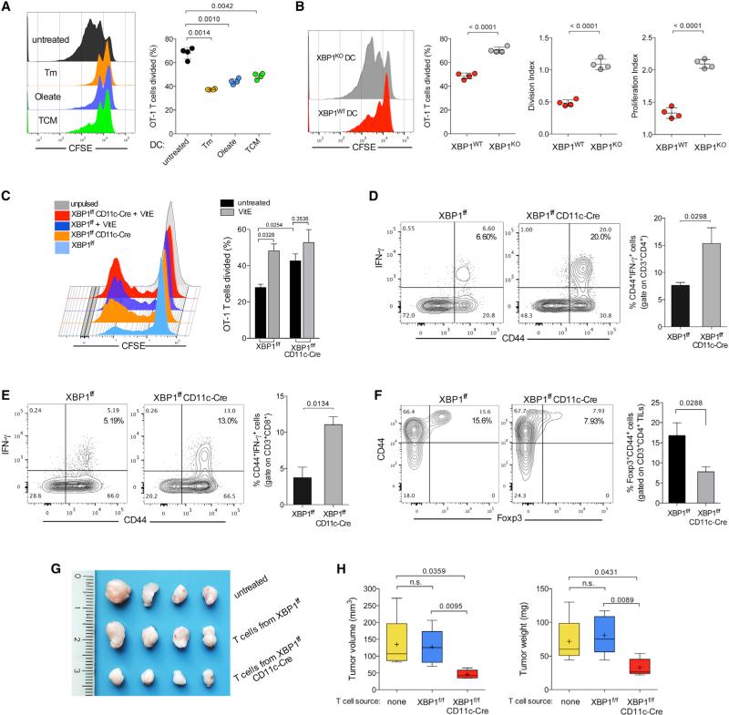
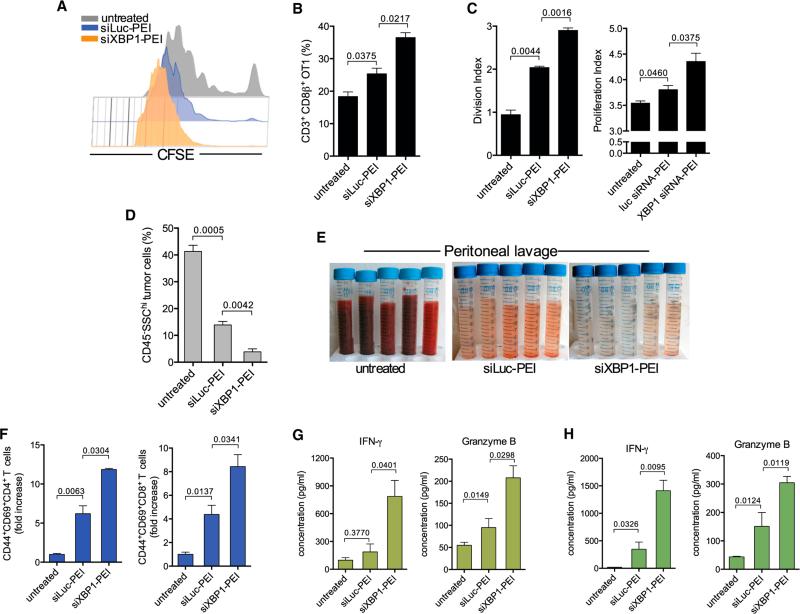
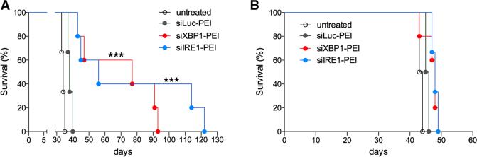
Dendritic cells (DCs) are required to initiate and sustain T cell-dependent anti-cancer immunity. However, tumors often evade immune control by crippling normal DC function. The endoplasmic reticulum (ER) stress response factor XBP1 promotes intrinsic tumor growth directly, but whether it also regulates the host anti-tumor immune response is not known. Here we show that constitutive activation of XBP1 in tumor-associated DCs (tDCs) drives ovarian cancer (OvCa) progression by blunting anti-tumor immunity. XBP1 activation, fueled by lipid peroxidation byproducts, induced a triglyceride biosynthetic program in tDCs leading to abnormal lipid accumulation and subsequent inhibition of tDC capacity to support anti-tumor T cells. Accordingly, DC-specific XBP1 deletion or selective nanoparticle-mediated XBP1 silencing in tDCs restored their immunostimulatory activity in situ and extended survival by evoking protective type 1 anti-tumor responses. Targeting the ER stress response should concomitantly inhibit tumor growth and enhance anti-cancer immunity, thus offering a unique approach to cancer immunotherapy.


Figures

Figure 1
Figure 1. XBP1 Activation in OvCa-Associated DCs
(A) Murine CD45+CD11c+MHC-II+CD11b+CD8αlow tDCs were isolated from advanced p53/K-ras-driven ovarian tumors or from malignant ascites of mice bearing aggressive ID8-Defb29/Vegf-A ovarian carcinoma for 4–5 weeks, and their identity as bona fide classical DCs was confirmed by quantifying Clec9A/DNGR-1 and Zbtb46 expression (see also Figure S1). CD45+CD11c+MHC-II+CD11b+CD8α splenic DCs (sDCs) were isolated from naive or tumor-bearing mice. Xbp1 splicing was evaluated using conventional RT-PCR. Xbp1u, unspliced form; Xbp1s, spliced form. (B–E) DCs were isolated from naive mice or from hosts bearing ID8-Defb29/Vegf-A OvCa for 3–4 weeks. (B, D, and E) Expression of the indicated transcripts was determined by RT-qPCR, and data were normalized to endogenous levels of Actb. (C) Western blot analysis of XBP1s expression in nuclear extracts obtained from the indicated DCs. Lamin B was used as loading control. (F) CD45+CD3 CD20 CD11c+DEC205+ tDCs were isolated from human patient ovarian tumors or metastatic OvCa ascites samples, and Xbp1 splicing was evaluated. (G) Expression of BiP and CHOP by all human tDC samples was analyzed. (H) The proportion of CD45+CD3+ tumor-infiltrating lymphocytes (TILs) was correlated with tDC CHOP mRNA expression in the same human specimen. r, Spearman's rank correlation coefficient.
Figure 2
Figure 2. OvCa Progression in Hosts Lacking XBP1 in DCs
(A) p53/K-ras-driven ovarian tumors were generated in hosts reconstituted with BM from either XBP1f/f (top) or XBP1f/f CD11c-Cre (bottom) donor mice. p53 deletion and oncogenic KRAS expression were induced via intrabursal injection of Cre-expressing adenovirus (ADV-Cre), as described in the Supplemental Experimental Procedures, and primary tumors were resected 7 weeks later. (B) Growth kinetics of p53/K-ras-driven tumors shown in (A). (C and D) Mice bearing primordial p53/K-ras-driven tumors for 28 days were injected i.p. with WT or XBP1-deficient (KO) BMDCs, and tumor growth was monitored over time until day 71. (E–H) Mice of the indicated genotypes were implanted with ID8-Defb29-VegfA OvCa cells via i.p. injection. (E and F) Peritoneal metastases evaluated 3–4 weeks after tumor implantation (n = 3 per group). (G) Malignant ascites generation in tumor-bearing mice expressed as percent weight gain due to progressive accumulation of peritoneal fluid. (H) Reduced splenomegaly in tumor-bearing mice deficient for XBP1 in CD11c+ DCs. (I and J) Survival in mice bearing aggressive ID8-Defb29-VegfA (I) or parental ID8 (J) tumors. Data are representative of at least two independent experiments with similar results using four to six mice per group. (K and L) ID8-Defb29-VegfA cancer cells were implanted in the flank alone or in combination with WT or XBP1-deficient (KO) BMDCs. Tumors were resected and measured 40 days later. *p < 0.05, **p < 0.001. Error bars represent SEM.
Figure 3
Figure 3. Lipid Peroxidation Byproducts Trigger ER Stress in DCs
(A) Intracellular lipid content in DC populations from naive or tumor-bearing mice (OvCa). (Left) Representative FACS analysis of lipid staining for DCs from the indicated sources. (Right) Lipid quantification expressed as mean fluorescence intensity (MFI) of Bodipy 493/503 staining. (B) Intracellular ROS levels in sDCs or in tDCs untreated or treated with vitamin E (VitE). Data are expressed as geometric MFI (gMFI) of DCFDA staining. (C) Intracellular 4-HNE-protein adducts in tDCs from human or mouse metastatic OvCa ascites samples. (D) Levels of 4-HNE-protein adducts in tDCs compared with control sDCs from tumor-bearing mice. (E) Dose-dependent increase in intracellular 4-HNE-protein adducts in sDCs exposed to ascites supernatants. (F) MS/MS spectrum on the doubly charged precursor at m/z 909.00, identifying a tryptic peptide with 4-HNE forming a Michael adduct on the Lys214 residue of the peptide from the 78 kDa glucose-regulated protein precursor (GRP78/BiP). (G) MS/MS spectrum on the triply charged precursor at m/z 643.32, identifying a tryptic peptide with 4-HNE forming a Michael adduct on the Lys82 residue of the peptide from the dnaJ homolog subfamily B member 11 precursor (ERdj3/Dnajb11). (H) Intracellular levels of 4-HNE protein adducts in sDCs exposed to cell-free ascites in the presence or absence of VitE or Hydralazine (Hlz) for 18 hr. (I and J) tDC were pretreated with VitE or Hlz and then incubated in normal media (gray bars) or in media supplemented with 25% cell-free ascites (green bars) for 18 hr. Expression of the indicated transcripts was assessed via RT-qPCR. Data are normalized to Actb expression in each sample. #p < 0.05 compared with tDC in ascites-free media. *p < 0.05 compared with ascites-exposed untreated tDC. Error bars represent SEM.
Figure 4
Figure 4. XBP1 Disrupts Lipid Homeostasis in OvCa-Associated DCs
(A and B) Downregulation of genes involved in the UPR/ER stress response (A) and lipid metabolism (B) in tDCs devoid of XBP1. WT is XBP1f/f tDC. XBP1def is XBP1f/f CD11c-Cre tDC (n = 3 per group). Black dots indicate genes involved in triglyceride biosynthesis. (C) Decreased intracellular lipid content in XBP1f/f CD11c-Cre tDCs (n = 3 mice per group) from mice bearing ID8-Defb29-VegfA tumors for 3 weeks revealed by Bodipy493/503 staining. (D) Electron micrographs (12,0003) showing large intracellular lipid bodies (red arrows) in XBP1-sufficient, but not XBP1-deficient, tDC. Black scale bars represent 2 μm. (E and F) Quantification of lipid bodies (E) and intracellular triacylglycerides (TAGs) (F) in tDCs sorted from mice bearing ID8-Defb29-VegfA ovarian tumors for 3–4 weeks. (G) tDCs were incubated in vitro with or without 25% cell-free OvCa ascites in the presence of 4μ8c, TOFA, Tiron, or VitE. Intracellular lipid content was assessed 24 hr later via Bodipy493/503 staining. Data are representative of three independent experiments with similar results. #p < 0.05 compared with control tDC incubated in the absence of cell-free ascites supernatants. *p < 0.05 compared with ascites-exposed tDC but left untreated. Error bars represent SEM.
Figure 5
Figure 5. XBP1 Promotes DC Malfunction in the OvCa Microenvironment
(A) WT BMDCs were left untreated or treated with tunicamycin (Tm), oleate, or TCM for 8 hr and then pulsed overnight with 100-μg/ml full-length OVA protein. CFSE-labeled OT-1 T cells were co-cultured with treated BMDCs, and proliferation was assessed by FACS 3 days later. (B) WT or XBP1-deficient (KO) BMDCs were incubated in 25% TCM for 8 hr, and the proliferation assay was repeated as described in (A). (C) CFSE-dilution analysis of OT-1 T cells co-cultured with OVA protein-pulsed tDCs isolated from the peritoneal cavity of XBP1f/f or XBP1f/f CD11c-Cre mice bearing ID8-Defb29-VegfA ovarian tumors for 4 weeks. tDCs were treated or untreated with vitamin E (VitE) during the OVA pulse. (D and E) Enhanced endogenous T cell activation at tumor sites in mice devoid of XBP1 in tDCs. Peritoneal wash samples from WT (XBP1f/f, black bars) or XBP1-deficient (XBP1f/f CD11c-Cre, gray bars) mice were collected 2–3 weeks after peritoneal implantation of ID8-Defb29-VegfA OvCa cells. Surface expression of CD44 and intracellular levels of tumoricidal IFN-γ were analyzed on CD3+CD4+ (D) or CD3+CD8+ (E) tumor-infiltrating T cells (TILs). (F) Proportion of T regulatory cells at tumor locations in mice of the indicated genotypes bearing metastatic ID8-Defb29-VegfA ovarian tumors for 3 weeks. In all cases, data are representative of two independent experiments using three to four mice per group. (G and H) Growth of solid ID8-Defb29-VegfA ovarian tumors in hosts adoptively transferred with splenic and draining lymph node T cells isolated from XBP1 f/f or XBP1f/f CD11c-Cre mice bearing metastatic tumors for 30 days. Error bars represent SEM.
Figure 6
Figure 6. Therapeutic Silencing of XBP1 Improves tDC Function and Evokes Anti-tumor Immunity
(A) Representative CFSE dilution of OT-1 T cells proliferating in vivo at tumor sites in OVA protein-pulsed untreated mice or after administration of immunostimulatory nanocomplexes carrying luciferase-matching (siLuc-PEI) or XBP1-specific (siXBP1-PEI) siRNA. (B) Proportion of OT-1 T cells in ascites of treated mice (n = 3 per group) 3 days after transfer. (C) Division and proliferation index of transferred OT-1 T cells shown in (B). (D–H) Enhanced anti-tumor immune responses in mice treated with DC-targeting, XBP1-silencing nanocomplexes. ID8-Defb29/Vegf-A tumor-bearing mice (n = 3 per group) were treated at days 8, 13, 18, and 23 post-tumor injection, and peritoneal lavage samples were analyzed at day 27. (D) Proportion of metastatic tumor cells (CD45 SSChi) found in the peritoneal cavity of treated mice. (E) Representative pictures of peritoneal lavages obtained from treated mice. (F) Proportion of antigen-experienced (CD44+), activated (CD69+) CD4+ (left), and CD8+ (right) T cells infiltrating tumor locations determined by FACS analyses (gated on CD3+ cells). (G and H) Representative ELISA-based analysis showing increased IFN-γ and Granzyme B secretion by peritoneal (G) and splenic (H) T cells isolated from mice treated with XBP1-silencing nanoparticles. Error bars represent SEM.
Figure 7
Figure 7. Therapeutic Effects of Targeting IRE1α/XBP1 Signaling in OvCa DCs
(A and B) Survival analysis in WT (A) or Rag2-deficient (B) OvCa-bearing mice (n = 6 per group) treated with nanocomplexes at days 12, 16, 20, 24, 28, and 32 after implantation of ID8-Defb29/Vegf-A cancer cells. Data are representative of two independent experiments with similar results. ***p < 0.001, log-rank test.

Comment in

References

    1. Acosta-Alvear D, Zhou Y, Blais A, Tsikitis M, Lents NH, Arias C, Lennon CJ, Kluger Y, Dynlacht BD. XBP1 controls diverse cell type- and condition-specific transcriptional regulatory networks. Mol. Cell. 2007;27:53–66. - PubMed
    1. Bollard CM, Gottschalk S, Leen AM, Weiss H, Straathof KC, Carrum G, Khalil M, Wu MF, Huls MH, Chang CC, et al. Complete responses of relapsed lymphoma following genetic modification of tumor-antigen presenting cells and T-lymphocyte transfer. Blood. 2007;110:2838–2845. - PMC - PubMed
    1. Caton ML, Smith-Raska MR, Reizis B. Notch-RBP-J signaling controls the homeostasis of CD8- dendritic cells in the spleen. J. Exp. Med. 2007;204:1653–1664. - PMC - PubMed
    1. Chen X, Iliopoulos D, Zhang Q, Tang Q, Greenblatt MB, Hatziapostolou M, Lim E, Tam WL, Ni M, Chen Y, et al. XBP1 promotes triple-negative breast cancer by controlling the HIF1α pathway. Nature. 2014;508:103–107. - PMC - PubMed
    1. Conejo-Garcia JR, Benencia F, Courreges MC, Kang E, Mohamed-Hadley A, Buckanovich RJ, Holtz DO, Jenkins A, Na H, Zhang L, et al. Tumor-infiltrating dendritic cell precursors recruited by a beta-defensin contribute to vasculogenesis under the influence of Vegf-A. Nat. Med. 2004;10:950–958. - PubMed

Publication types

Associated data