. 2015 Jun 23;11(11):1686-93.
doi: 10.1016/j.celrep.2015.05.023. Epub 2015 Jun 11.

Iron Toxicity in the Retina Requires Alu RNA and the NLRP3 Inflammasome

Affiliations

Iron Toxicity in the Retina Requires Alu RNA and the NLRP3 Inflammasome

Bradley D Gelfand et al. Cell Rep. .

Abstract

Excess iron induces tissue damage and is implicated in age-related macular degeneration (AMD). Iron toxicity is widely attributed to hydroxyl radical formation through Fenton's reaction. We report that excess iron, but not other Fenton catalytic metals, induces activation of the NLRP3 inflammasome, a pathway also implicated in AMD. Additionally, iron-induced degeneration of the retinal pigmented epithelium (RPE) is suppressed in mice lacking inflammasome components caspase-1/11 or Nlrp3 or by inhibition of caspase-1. Iron overload increases abundance of RNAs transcribed from short interspersed nuclear elements (SINEs): Alu RNAs and the rodent equivalent B1 and B2 RNAs, which are inflammasome agonists. Targeting Alu or B2 RNA prevents iron-induced inflammasome activation and RPE degeneration. Iron-induced SINE RNA accumulation is due to suppression of DICER1 via sequestration of the co-factor poly(C)-binding protein 2 (PCBP2). These findings reveal an unexpected mechanism of iron toxicity, with implications for AMD and neurodegenerative diseases associated with excess iron.


Figures

Figure 1
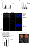
Figure 1. Iron toxicity depends on the NLRP3 inflammasome
(A) Immuno-labeling of Nlrp3 (yellow signal) and nuclei (DAPI, blue signal) in the RPE/choroid of 6-month old wild-type (top) or Cp−/−Heph−/− mice (middle). Bottom row depicts the same area from a serial section of Cp−/−Heph−/− immunolabeled with isotype goat IgG. Representative of N=3 mice. Scale bar denotes 20 μm. (B) Fundus (top) and ZO-1 staining of RPE flat mount preparations (bottom) of wild-type mice 7 days after injection of 1μL Fe(III) at indicated concentrations. Blue arrows denote injection site. (C and D) Western blotting of pro- and active forms of Caspase-1 in ARPE-19 cells and wild-type mouse retina and RPE/choroid (pooled from 4 eyes) treated with indicated doses of Fe(III). Fundus and ZO-1 staining of RPE flat mount preparations following delivery of Fe(III) into the subretinal space of mice lacking inflammasome components Caspase-1/11 (E) and Nlrp3 (F). (G) Fundus and ZO-1 staining of RPE flat mount preparations of wild-type mice that received Caspase-1 peptide inhibitor preceding delivery of Fe(III) into the subretinal space. (H, I) Fundus and ZO-1 staining of RPE flat mount preparations following administration of Cr(VI), Cu(I) or Zn(I) into the subretinal space of wild-type mice (H) and in mice lacking inflammasome components Caspase-1/11 (I). Indicated doses represent the minimum concentration of metals required to consistently induce RPE degeneration. (B-I) Scale bars denote 50 μm. Fundus images representative of at least 4 replicates.
Figure 2
Figure 2. Iron toxicity depends on SINE RNA induction
(A) Densitometry of Alu RNA northern blotting and real-time qPCR of DICER1 mRNA in human ARPE-19 cells exposed to iron overload for 4 days at indicated doses. (B) Densitometry of B1 and B2 RNAs northern blotting and real-time qPCR of Dicer1 mRNA RPE/choroid lysates from wild-type C57BL6/J mice 7 days after subretinal injection with iron. (C) Fluorescent in situ hybridization of B1 and B2 RNAs in retinal cross-sections of 6-month old Cp−/−Heph−/− (top row) or wild-type mice (bottom row). Scale bar denotes 50 μm. (D) Western blotting of pro- and active forms of Caspase-1 in ARPE-19 cells exposed to Fe(III) after treatment with either scrambled or Alu RNA-targeted antisense oligonucleotides. (E) Representative fundus photographs and ZO-1 immunostained RPE flat mounts of wild-type mice treated with a cell-permeable antisense oligonucleotide targeting B2 RNA (and scrambled control) 1 day prior to subretinal injection of 10 nM Fe(III). Images were acquired 6 days after Fe(III) administration. Blue arrows denote injection site. Scale bar denotes 50 μm. For all panels, N=3-6, *P < 0.05, Error bars denote SEM.
Figure 3
Figure 3. Iron overload enhances stability of Alu RNA
(A) Processing of a synthetic biotin-labeled Alu RNA transiently transfected in ARPE-19 exposed to 1 mM Fe(III). X-axis denotes time following 2 h RNA loading period. (B) Quantity of endogenous Alu RNA accumulated during a 4 hour modified nucleotide doping pulse (4 h) or after washout (20 h) in ARPE-19 cells exposed to 1 mM Fe(III). Alu RNAs were quantified by q-PCR of size-separated RNA samples, in which RNAs 100-800 nt were isolated after poly-acrylamide gel electrophoresis. (C) Northern blotting of Alu RNA of human ARPE-19 cells treated with control or DICER1-targeted antisense oligonucleotides, and exposed to indicated doses of Fe(III). (D) Polyacrylamide gel separated Alu RNA after incubation with recombinant DICER1 in the presence of indicated quantities of Fe(III).
Figure 4
Figure 4. Iron sensitivity of DICER1 processing of Alu RNAs is mediated by PCBP2
(A) Western blotting of streptavidin-mediated pull-down and whole cell lysates from biotin-Alu or biotin-tRNA transfected human ARPE-19 cells that were exposed to 1 mM Fe(III). (B) Northern blotting of native Alu and 5S RNAs in human ARPE-19 exposed to 1 mM Fe(III) following immuno-precipitation with anti-PCBP2 antibody or in whole cell lysates. (C) DICER1-mediated Alu RNA cleavage quantified from in vitro dicing reactions containing recombinant human DICER1, recombinant human PCBP2, and 100 μM Fe(III) where indicated. For all panels, N=3-6, *P < 0.05, Error bars denote SEM. (D) Proposed model of iron overload-induced retinal toxicity, involving the sequestration of PCBP2 from Alu RNA/DICER1 complexes, leading to an accumulation of Alu RNAs, NLRP3 inflammasome activation and retinal degeneration.

References

    1. Anderson OA, Finkelstein A, Shima DT. A2E induces IL-1ss production in retinal pigment epithelial cells via the NLRP3 inflammasome. PloS one. 2013;8:e67263. - PMC - PubMed
    1. Ayton S, Lei P. Nigral iron elevation is an invariable feature of Parkinson's disease and is a sufficient cause of neurodegeneration. BioMed research international. 2014;2014:581256. - PMC - PubMed
    1. Blyn LB, Swiderek KM, Richards O, Stahl DC, Semler BL, Ehrenfeld E. Poly(rC) binding protein 2 binds to stem-loop IV of the poliovirus RNA 5′ noncoding region: identification by automated liquid chromatography-tandem mass spectrometry. Proceedings of the National Academy of Sciences of the United States of America. 1996;93:11115–11120. - PMC - PubMed
    1. Blyn LB, Towner JS, Semler BL, Ehrenfeld E. Requirement of poly(rC) binding protein 2 for translation of poliovirus RNA. Journal of virology. 1997;71:6243–6246. - PMC - PubMed
    1. Chan C, Shen D, Wang Y, Chu X, Abu-Asab M, Tuo J. Inflammasomes in human eyes with AMD and mouse retinas with focal retinal degeneration; ARVO Annual Meeting; Seattle, WA. 2013. p. 315.

Publication types

MeSH terms

LinkOut - more resources