. 2015 Jun 23;11(11):1714-26.
doi: 10.1016/j.celrep.2015.05.032. Epub 2015 Jun 11.

In Vivo RNAi Screening Identifies MDA5 as a Significant Contributor to the Cellular Defense against Influenza A Virus

Affiliations

In Vivo RNAi Screening Identifies MDA5 as a Significant Contributor to the Cellular Defense against Influenza A Virus

Asiel A Benitez et al. Cell Rep. .

Abstract

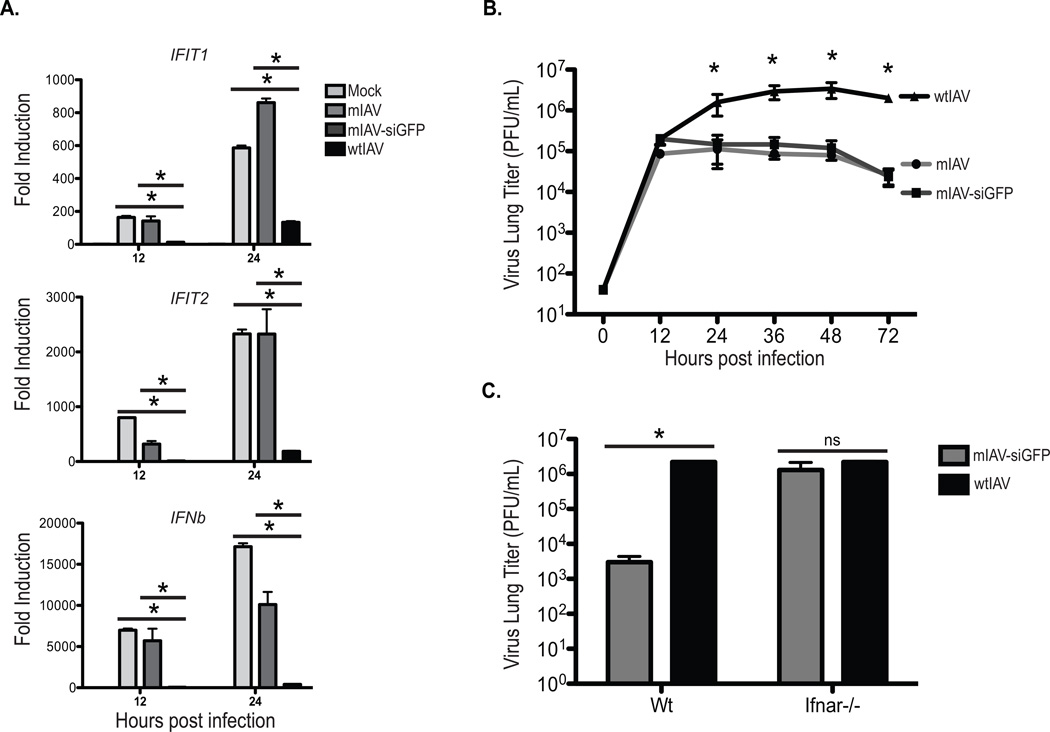
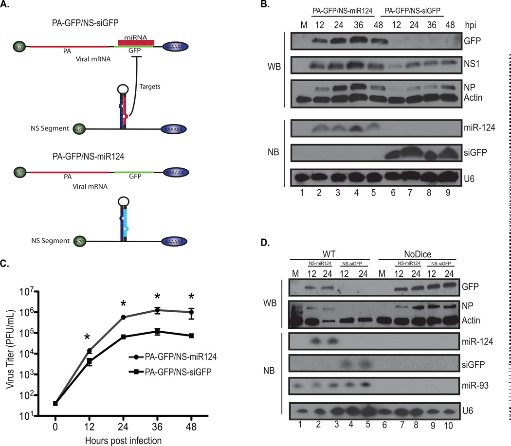
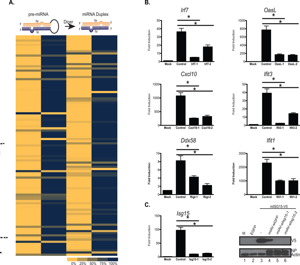
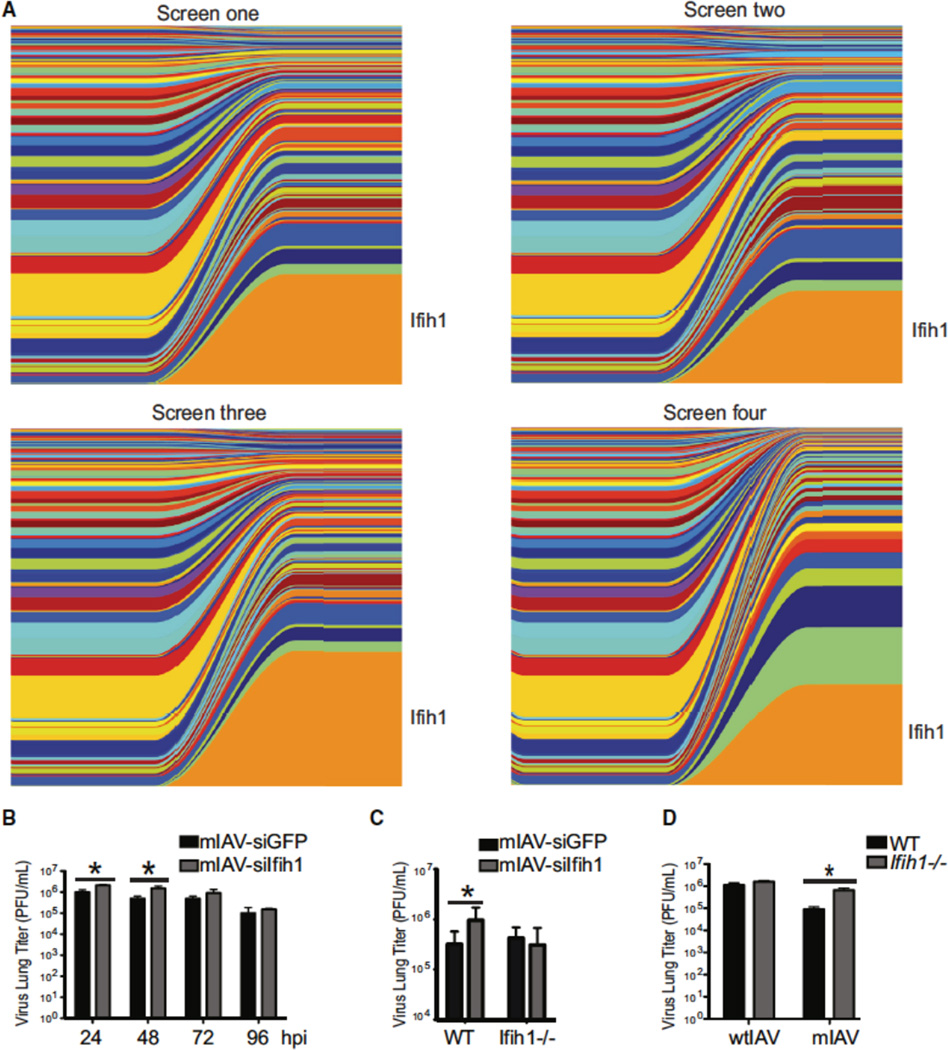
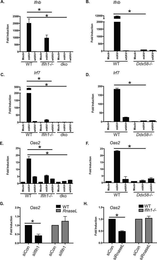
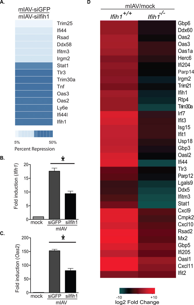
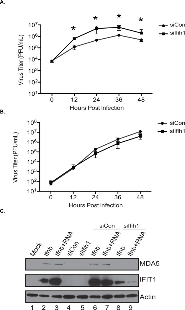
Responding to an influenza A virus (IAV) infection demands an effective intrinsic cellular defense strategy to slow replication. To identify contributing host factors to this defense, we exploited the host microRNA pathway to perform an in vivo RNAi screen. To this end, IAV, lacking a functional NS1 antagonist, was engineered to encode individual siRNAs against antiviral host genes in an effort to rescue attenuation. This screening platform resulted in the enrichment of strains targeting virus-activated transcription factors, specific antiviral effectors, and intracellular pattern recognition receptors (PRRs). Interestingly, in addition to RIG-I, the PRR for IAV, a virus with the capacity to silence MDA5 also emerged as a dominant strain in wild-type, but not in MDA5-deficient mice. Transcriptional profiling of infected knockout cells confirmed RIG-I to be the primary PRR for IAV but implicated MDA5 as a significant contributor to the cellular defense against influenza A virus.


Figures

Figure 1
Figure 1. Virus-encoded siRNAs do not impose further attenuation on mIAV
(A) Quantitative RT-PCR (qPCR) of IFIT1, IFIT2, and IFNb in A549 cells either mock treated or infected (MOI=0.1) with mIAV, mIAV-siGFP, or wtIAV. Error bars, SD, *p<0.05. (B) Multi-cycle growth curves in A549s infected (MOI=0.01) with mIAV, mIAV-siGFP, or wtIAV. Error bars, SD, *p<0.05. (C) Virus lung titers of wild type or Ifnar1−/− B6 mice infected with either the mIAV-siGFP, or wtIAV viruses. Mice were infected with 1 × 10⁶ pfu of each virus and lungs were harvested 24 hpi. Error bars, SD, *p<0.05.
Figure 2
Figure 2. The kinetics of virus-derived siRNAs are sufficient to knockdown virus or IFN-induced genes
(A) Schematic depicting a self-targeted virus (PA-GFP/ NS-siGFP) and the control virus (PA-GFP/NS-miR124). Shown is the PA segment encoding GFP and the NS segment encoding an siRNA targeting GFP (siGFP) or a non-targeting small RNA (miR-124). (B) Multi-cycle growth curves in A549s infected (MOI=0.01) with viruses in (A). Error bars, SD, *p<0.05. (C) Top three panels depict western blots of whole cell extract derived from A549 cells either mock treated or infected (MOI=0.01) with PA-GFP/NS-siGFP or PA-GFP/NS-miR124. Immunoblots were probed for GFP, IAV NP, IAV NS1, and Actin, which was used as a loading control. Bottom three panels depict northern blots of total RNA derived from A549s. Northern blots were probed for small RNA expression of microRNA-124 (miR-124) the GFP targeting siRNA (siGFP) and the host splicing RNA, U6, which was used as a loading control. (D) Top two panels depict western blots of whole cell extract derived from wild type 293T cells or NoDice cells (293T cells lacking Dicer). Cells were either mock treated or infected (MOI=0.01) with PA-GFP/NS-siGFP or PA-GFP/NS-miR124 at the time points indicated. Immunoblots were probed for GFP, IAV NP, and Actin as in (C). Bottom four panels depict northern blots of total RNA derived from the conditions above. Northern blots were probed for small RNA expression of miR-124, siGFP, miR-93, and U6.
Figure 3
Figure 3. Constructing a library of siRNAs targeting virus or IFN induced genes
(A) Schematic depicting the structure of pre- and mature miRNA duplex with the 5p (orange) and 3p (blue) strands highlighted. Beneath this is a heat map of relative read counts derived from small RNAseq analysis from small RNAs derived from A549s infected with the IAV library. Results are shown as the proportion of the 5p and 3p produced for each siRNA. (B) Quantitative RT-PCR (qPCR) of mRNA derived from mouse embryonic fibroblasts mock treated or infected (MOI=3) with viruses targeting IRF7, CXCL10, RIG-I, OASL, IFIT3, and IFIT1. Each qPCR was generated to determine the expression profile of the cognate target. Each virus set included two unique siRNA hairpins (−1 or −2). Error bars, SD, *p<0.05. (C) RNA and protein analysis of ISG15 from MEFs mock treated or infected (MOI=3) with the corresponding siISG15 viruses (Isg15-1 or Isg15-2). Top panel depict qPCR analysis for ISG15 as described in (B), bottom panel depicts a western blot of whole cell extract derived from 293 cells transfected with epitope tagged (V5) murine ISG15 and infected with the aforementioned viruses. Immunoblots were probed form V5, IAV NP, and Actin.
Figure 4
Figure 4. Infection with the library in mice selects for a virus targeting Ifih1
(A) Graphical depiction of siRNA composition derived from deep sequencing data of mice infected with 1 × 10⁴ pfu of the pooled library. Left side of each graph represents the proportion of unique viral strains, as defined by their siRNA. Right side of each graph depicts in vivo selection dynamics following four days of infection. (B) Levels of virus replication from mice infected with 1 × 10⁶ pfu of mIAV-siGFP or -siIfih1 and assessed at the indicated time points. Error bars, SD, *p<0.05. (C) Lung titers from wild-type or Ifih1−/− mice infected with 1 × 10⁶ pfu of mIAV-siGFP or -siIfih1 as measured 24 hours post infection. Error bars, SD, *p<0.05. (D) Lung titers from wild-type mice or Ifih1−/− mice infected with 1 × 10⁴ pfu of wtIAV or mIAV measured 48 hours post infection. Error bars, SD, *p<0.05.
Figure 5
Figure 5. MDA5 is a non-redundant pattern recognition receptor for IAV
(A) qPCR of Ifnb derived from wild-type, Ifih1−/−, or Ddx58/Ifih1−/− (dko) murine fibroblast cells either mock treated or infected with mIAV or wtIAV (MOI=2). Error bars, SD, *p<0.05. (B) qPCR of Ifnb derived from wild-type, or Ddx58−/− murine fibroblast cells either mock treated or infected with mIAV or wtIAV (MOI=2). Error bars, SD, *p<0.05. (C) qPCR as described in (A) for Irf7. (D) qPCR as described in (B) for Irf7. (C) qPCR as described in (A) for Oas2. (D) qPCR as described in (B) for Oas2. F) qPCR of Oas2 derived from wild-type, or RnaseL−/− murine fibroblast cells either mock treated or infected with mIAV (MOI=2). Error bars, SD, *p<0.05. (G) qPCR of Oas2 derived from wild-type, or Ifih1−/− murine fibroblast cells either mock treated or infected with mIAV (MOI=2). Error bars, SD, *p<0.05.
Figure 6
Figure 6. Infection with mIAV-siIfih1 results in an altered ISG profile
(A) Transcriptional profiling of murine fibroblasts infected (MOI=2) with mIAV-siGFP or -siIfih1 as determined by mRNAseq. The heat map depicts fold repression of mIAV-siGFP-induced genes in response to mIAV-siIfih1. (B) qPCR of Ifih1 derived from samples described in (A). Error bars, SD, *p<0.05. (C) qPCR as described in (B) for Oas2 (D) Heat map depicting expression changes in a subset of antiviral genes in wild-type or Ifih1−/− murine fibroblasts infected with mIAV (MOI=2) for 12 hours. Values represent log2 fold induction of infected over uninfected cells.
Figure 7
Figure 7. MDA5 function of IAV detection is conserved between mouse and human cells
(A) Multi-cycle growth curves performed in A549 cells treated with a scrambled (scbl) or IFIH1- specific siRNAs. Cells were infected (MOI=0.01) with mIAV and supernatants collected and plaqued at the indicated time points. Error bars, SD, *p<0.05. (B) Multi-cycle growth curves as described in (A) using wild type IAV in place of mIAV. (C) Western Blot of whole cell extract derived from A549 cells treated as in (A) and subsequently transfected with IAV RNA and/or administered IFNβ. Immunoblots were probed for MDA5, IFIT1, and Actin.

References

    1. Andrejeva J, Childs KS, Young DF, Carlos TS, Stock N, Goodbourn S, Randall RE. The V proteins of paramyxoviruses bind the IFN-inducible RNA helicase, mda-5, and inhibit its activation of the IFN-beta promoter. Proc Natl Acad Sci U S A. 2004;101:17264–17269. - PMC - PubMed
    1. Bartel DP. MicroRNAs: target recognition and regulatory functions. Cell. 2009;136:215–233. - PMC - PubMed
    1. Bogerd HP, Whisnant AW, Kennedy EM, Flores O, Cullen BR. Derivation and characterization of Dicer- and microRNA-deficient human cells. RNA. 2014;20:923–937. - PMC - PubMed
    1. Brass AL, Huang IC, Benita Y, John SP, Krishnan MN, Feeley EM, Ryan BJ, Weyer JL, van der Weyden L, Fikrig E, et al. The IFITM proteins mediate cellular resistance to influenza A H1N1 virus, West Nile virus, and dengue virus. Cell. 2009;139:1243–1254. - PMC - PubMed
    1. Childs K, Stock N, Ross C, Andrejeva J, Hilton L, Skinner M, Randall R, Goodbourn S. mda-5, but not RIG-I, is a common target for paramyxovirus V proteins. Virology. 2007;359:190–200. - PubMed

Publication types

Substances