

Spontaneous epidural hematoma of the spine associated with oral anticoagulants: 3 Case Studies
- PMID: 26074484
- PMCID: PMC4529632
- DOI: 10.1016/j.ijscr.2015.05.022
Spontaneous epidural hematoma of the spine associated with oral anticoagulants: 3 Case Studies
Abstract
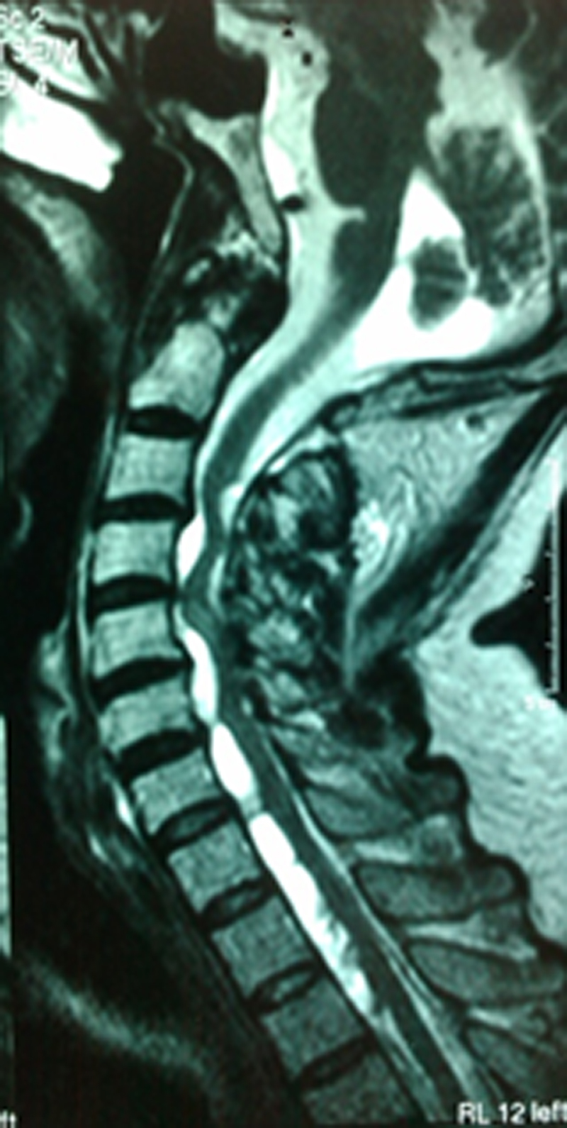
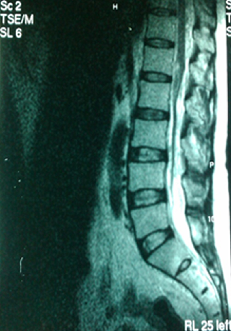
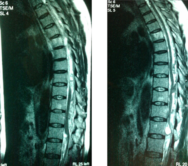
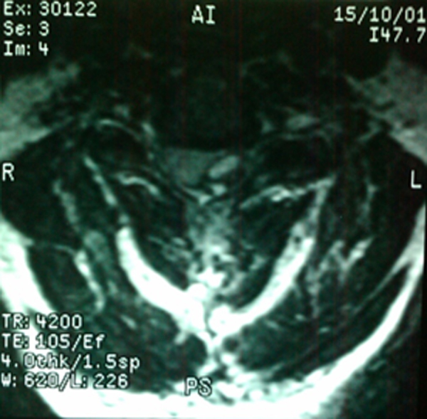
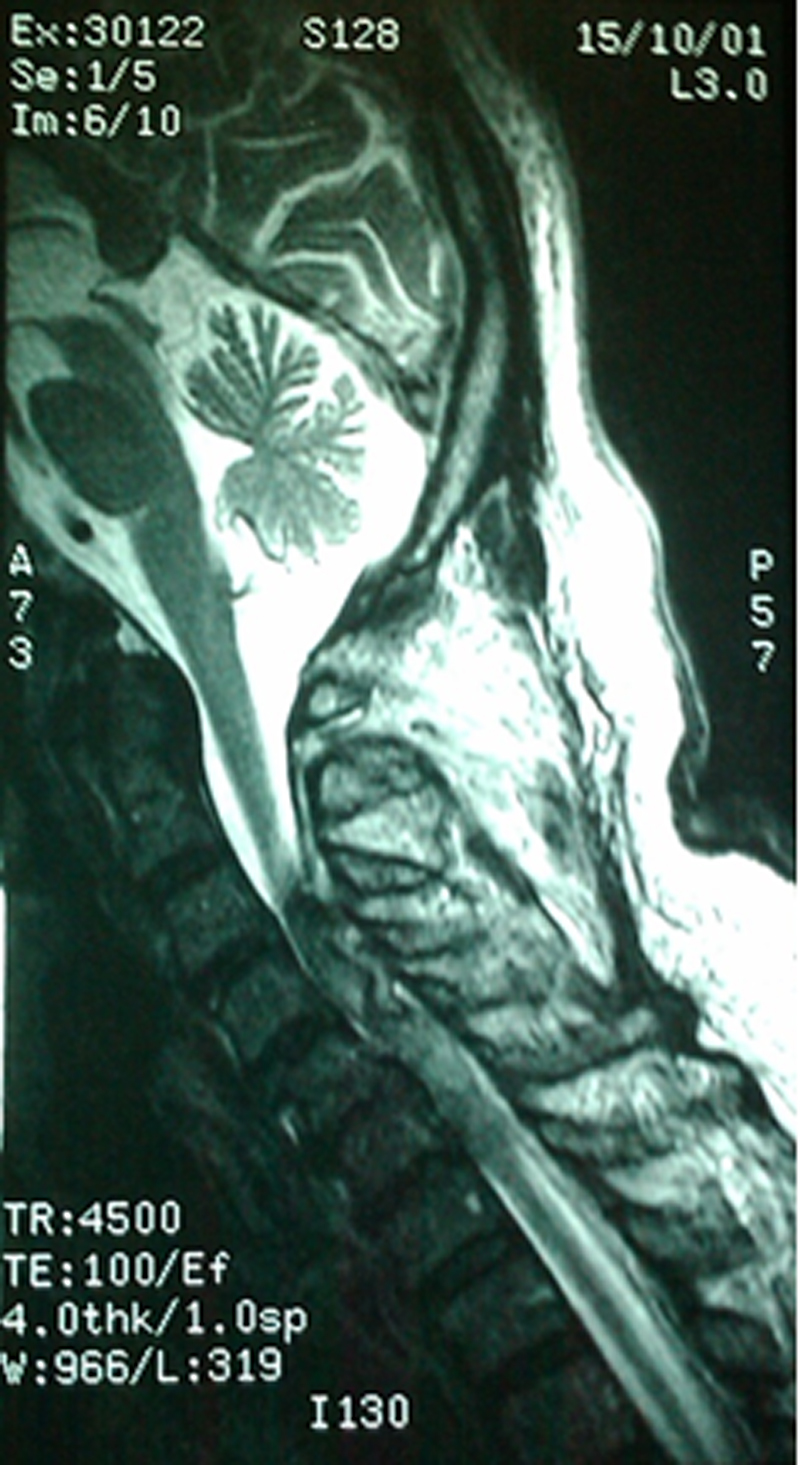
Introduction: Spontaneous epidural hematoma of the spine (SEHS) is an extremely rare entity. Patients known to have thrombophilia or on anticoagulant drugs are the most affected. It is generally caused by a rupture of the postero-internal vertebral venous plexus secondary to minor barotrauma such as cough, peridural catheter insertion... Early diagnosis and treatment showed to have the best outcome.
Cases report: We report 3 cases of spontaneous epidural hematoma in patients treated by acenocoumarol, which occurred without any provocative factor. All 3 patients were treated with decompressive laminectomy.
Discussion: We described the MRI findings and discussed the spontaneity of the entity. Our present case studies and the review of the literature showed that early diagnosis and management of SEHS can lead to improvement of the neurological state and avoid definitive motor and sensitive deficit.
Conclusion: This rare entity should be suspected in every acute medullary syndrome with spinal pain, motor and/or sensory deficit. In order to decrease the sequelae, neurologically unstable patients should benefit from early diagnosis and urgent surgical decompression.
Keywords: Acenocoumarol; Oral anticoagulants; Spontaneous epidural hematoma of the spine.
Copyright © 2015 The Authors. Published by Elsevier Ltd.. All rights reserved.
Figures
References
-
- Jackson R. Case of spinal apoplexy. Lancet. 1869;2:5.
-
- Kreppel D., Antoniadis G., Seeling W. Spinal hematoma: a literature survey with a meta-analysis of 613 patients. Neurosurg. Rev. 2003;26:1–49. - PubMed
-
- Beatty R.M., Winston K.R. Spontaneous cervical epidural hematoma. A consideration of etiology. J. Neurosurg. 1984;61:143–148. - PubMed
-
- Groen R.J.M., Ponssen H. The spontaneousspinal epidural hematoma. A study of the etiology. J. Neurol. Sci. 1990;98:121–138. - PubMed
-
- Nuti C., Fotso M.J., Duhel R., Hatem B., Dumas B., Brunon J. Hématomes épiduraux non traumatiques du rachis. Présentation de 20 cas. Revue de la littérature et étude des aspects évolutifs. Neurochirurgie. 2003;49:563–570. - PubMed
LinkOut - more resources
Full Text Sources
Other Literature Sources
