. 2015 May 29:9:184.
doi: 10.3389/fnins.2015.00184. eCollection 2015.

The fluorescent pentameric oligothiophene pFTAA identifies filamentous tau in live neurons cultured from adult P301S tau mice

Affiliations

The fluorescent pentameric oligothiophene pFTAA identifies filamentous tau in live neurons cultured from adult P301S tau mice

Jack Brelstaff et al. Front Neurosci. .

Abstract

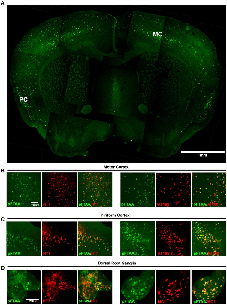
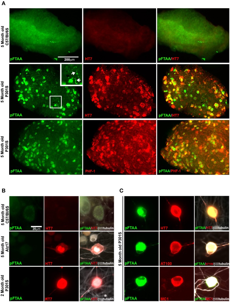
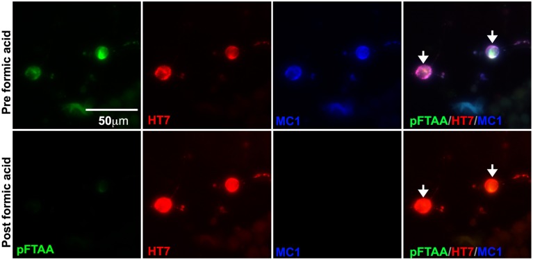
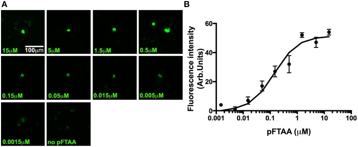
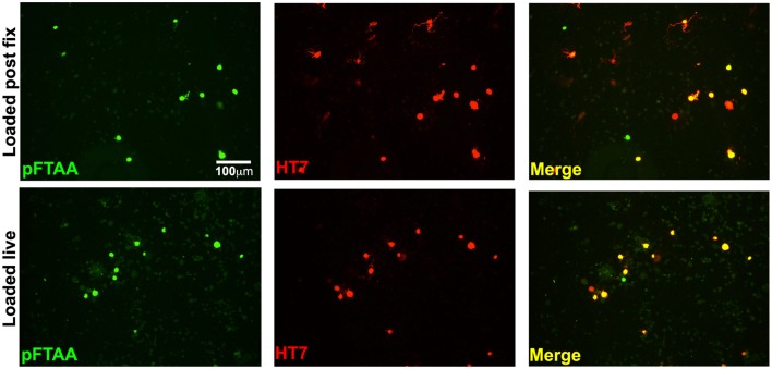
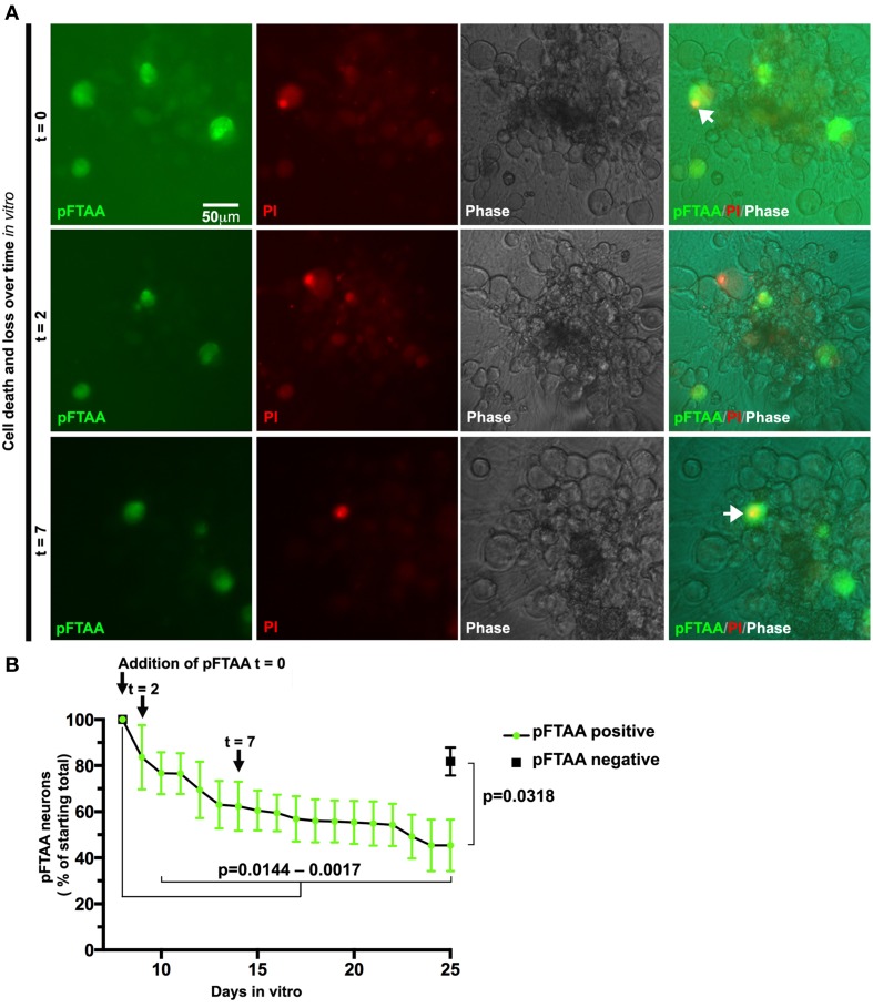
Identification of fluorescent dyes that label the filamentous protein aggregates characteristic of neurodegenerative disease, such as β-amyloid and tau in Alzheimer's disease, in a live cell culture system has previously been a major hurdle. Here we show that pentameric formyl thiophene acetic acid (pFTAA) fulfills this function in living neurons cultured from adult P301S tau transgenic mice. Injection of pFTAA into 5-month-old P301S tau mice detected cortical and DRG neurons immunoreactive for AT100, an antibody that identifies solely filamentous tau, or MC1, an antibody that identifies a conformational change in tau that is commensurate with neurofibrillary tangle formation in Alzheimer's disease brains. In fixed cultures of dorsal root ganglion (DRG) neurons, pFTAA binding, which also identified AT100 or MC1+ve neurons, followed a single, saturable binding curve with a half saturation constant of 0.14 μM, the first reported measurement of a binding affinity of a beta-sheet reactive dye to primary neurons harboring filamentous tau. Treatment with formic acid, which solubilizes filamentous tau, extracted pFTAA, and prevented the re-binding of pFTAA and MC1 without perturbing expression of soluble tau, detected using an anti-human tau (HT7) antibody. In live cultures, pFTAA only identified DRG neurons that, after fixation, were AT100/MC1+ve, confirming that these forms of tau pre-exist in live neurons. The utility of pFTAA to discriminate between living neurons containing filamentous tau from other neurons is demonstrated by showing that more pFTAA+ve neurons die than pFTAA-ve neurons over 25 days. Since pFTAA identifies fibrillar tau and other misfolded proteins in living neurons in culture and in animal models of several neurodegenerative diseases, as well as in human brains, it will have considerable application in sorting out disease mechanisms and in identifying disease-modifying drugs that will ultimately help establish the mechanisms of neurodegeneration in human neurodegenerative diseases.

Keywords: Alzheimer's disease; dorsal root ganglion neurons; fluorescent vital fibrillar tau dye; frontotemporal dementia (FTD); hyperphosphorylated tau; neurofibrillary tangles; transgenic P301S tau mouse.


Figures

Figure 1
Figure 1
Labeling of tau inclusions in the brain and DRG of 5-month-old P301S transgenic mouse by pFTAA. A P301S tau transgenic mouse injected with 150 nmole pFTAA via the tail vein was perfused after 48 h. (A) A composite image of a 30 μm coronal brain section at the level of the anterior commissure (5x objective) showing the general staining pattern of pFTAA+ve neurons; staining is found almost exclusively in the cortex. MC, motor M1 cortex, PC, piriform cortex. (B) motor M1 cortex and (C) piriform cortex stained with pFTAA (green) and immunolabeled with anti-human tau antibody HT7, or anti-fibrillar phospho-tau-specific antibody AT100 (red). (D) whole mount of a DRG stained with pFTAA in vivo (green) and immunolabeled with anti-human tau antibody HT7, or the conformational anti-tau antibody MC1 (red). Note that due to poorer penetration of the antibodies compared to pFTAA, some pFTAA+ve neurons that appear HT7-ve at the exposure shown are co-stained by the antibodies upon increased exposure. Scale bar, 1 mm (A) 100 μm (B,C), 250 μm (D).
Figure 2
Figure 2
pFTAA selectively labels fibrillar tau in DRG neurons in intact DRG ex vivo and cultured dissociated DRG neurons from 5-month-old P301S tau transgenic mice. (A) Intact DRG from 5-month-old C57BL/6 OlaHsd (wt, top row) or P301S tau mice (middle row) were incubated with 15 μM pFTAA at 4°C for 15 h, then fixed in 95% ethanol, rehydrated, and immunolabeling (red) with the HT7 antibody. Bottom row shows immunolabeling with the PHF-1 antibody (which recognizes phospho-serines396/404). Magnification box identifies pFTAA+ve threads visible in the axons of whole ganglia (arrows). Non-specific signal in wt intact ganglia is a-cellular debris carried over because of re-use of the dye. (B) DRG neurons were cultured from 5-month-old C57BL/6 OlaHsd wild type mice, or Alz17 mice transgenic for the longest isoform of human tau (2N4R), or 2-month-old P301S tau mice for 2 days. After ethanol fixation, cultures were labeled with pFTAA and immunolabeled with HT7. Note absence of pFTAA staining but presence of human tau in the neurons from Alz17 and 2-month-old P301S tau mice. (C) Dissociated DRG neurons from 5-month-old P301S tau mice were labeled after ethanol fixation by pFTAA (green), as well as for AT100 or MC1 (red). Images are representative of 133–224 pFTAA+ve cells across 3 biological replicates. Scale bar, 200 μm in (A) 20 μm in (B,C).
Figure 3
Figure 3
Formic acid treatment solubilizes fibrillar tau, but not souble tau, and eliminates the binding of pFTAA. DRG neurons cultured from 5-month-old P301S tau mice for 2 days were fixed with 95% ethanol. Top row shows pFTAA+ve neurons immunolabeled with HT7 (red) and MC1 (blue) antibodies. Bottom row shows the same set of neurons re-stained with pFTAA and antibodies after a 15 min treatment with 90% formic acid. Note that formic acid treatment prevented the re-binding of pFTAA and the MC1 antibody whereas the interaction with HT7, which binds to soluble and insoluble tau, was restored. Scale bar, 50 μm.
Figure 4
Figure 4
A dose response of pFTAA demonstrates a single, homogeneous binding site to neurons expressing filamentous tau. pFTAA was added at the concentrations indicated to ethanol-fixed DRG neurons cultured from 5-month-old P301S tau mice for 2 days. (A) Representative images of pFTAA binding intensities at the concentrations indicated. (B) Quantification of signal intensity. To determine the relationship between the concentration of pFTAA and fluorescence intensity, the fluorescence intensity values from 15 neurons per dose were averaged (circles) and the results were submitted to a least square curve fitting analysis according to an equation that predicts a single, saturable binding site (line) from which a half saturation constant of 0.142 μM (R2 = 0.983) was derived. Error bars indicate sem values. Scale bar, 100 μm.
Figure 5
Figure 5
Labeling of insoluble tau is not an artifact of fixation. DRG neurons cultured from 5-month-old P301S tau mice for 2 days were either incubated with 3 μM pFTAA for 30 min at room temperature before fixation in 95% ethanol, or fixed before labeling with pFTAA using the same solution and conditions. Cultures were immunolabeled with the HT7 antibody (red). Loading of pFTAA into either live or fixed dissociated DRG neurons produced equivalent signal and co-localization with HT7. Note that some HT7+ve neurons are not labeled with pFTAA because HT7 neurons with insoluble tau only represent about 44% of all the HT7+ve neurons. Scale bar, 100 μm.
Figure 6
Figure 6
Slow death of defined DRG neurons expressing fibrillar tau can be followed continuously after labeling with pFTAA. DRG neurons were cultured from 5-month-old P301S tau mice for 7 days before labeling with 3 μM pFTAA, (t = time in days after addition of pFTAA). Medium was replaced with PI-containing medium without pFTAA every 3 days. (A) Images of the same set of neurons at 0, 2, and 7 days after addition of pFTAA showing appearance of PI+ve neurons and their subsequent disappearance from the cultures. (B) The loss of pFTAA+ve neurons as a percentage of total neurons originally labeled is 45% after 25 days in vitro. The loss of pFTAA+ve neurons becomes significant at 10 days in vitro (p = 0.0144) and continues to increase until 25 days in vitro (p = 0.0017). Black square denotes percentage survival of pFTAA-ve neurons in the same culture after 25 days (mean ± sem from the three biological replicates, p = 0.0318, paired t-test). Scale bar, 50 μm.

Comment in

References

    1. Allen B., Ingram E., Takao M., Smith M. J., Jakes R., Virdee K., et al. . (2002). Abundant tau filaments and nonapoptotic neurodegeneration in transgenic mice expressing human P301S tau protein. J. Neurosci. 22, 9340–9351. - PMC - PubMed
    1. Aslund A., Sigurdson C. J., Klingstedt T., Grathwohl S., Bolmont T., Dickstein D. L., et al. . (2009). Novel pentameric thiophene derivatives for in vitro and in vivo optical imaging of a plethora of protein aggregates in cerebral amyloidoses. ACS Chem. Biol. 4, 673–684. 10.1021/cb900112v - DOI - PMC - PubMed
    1. Bandyopadhyay B., Li G., Yin H., Kuret J. (2007). Tau aggregation and toxicity in a cell culture model of tauopathy. J. Biol. Chem. 282, 16454–16464. 10.1074/jbc.M700192200 - DOI - PubMed
    1. Binder L. I., Frankfurter A., Rebhun L. I. (1985). The distribution of tau in the mammalian central nervous system. J. Cell Biol. 101, 1371–1378. 10.1083/jcb.101.4.1371 - DOI - PMC - PubMed
    1. Bing G., Nguyen X. V., Liu M., Markesbery W. R., Sun A. (2006). Biophysical and biochemical characterization of the intrinsic fluorescence from neurofibrillary tangles. Neurobiol. Aging 27, 823–830. 10.1016/j.neurobiolaging.2005.04.005 - DOI - PubMed