Review
. 2015 May 29:9:202.
doi: 10.3389/fncel.2015.00202. eCollection 2015.

Cellular and Deafness Mechanisms Underlying Connexin Mutation-Induced Hearing Loss - A Common Hereditary Deafness

Affiliations
Review

Cellular and Deafness Mechanisms Underlying Connexin Mutation-Induced Hearing Loss - A Common Hereditary Deafness

Jeffrey C Wingard et al. Front Cell Neurosci. .

Abstract

Hearing loss due to mutations in the connexin gene family, which encodes gap junctional proteins, is a common form of hereditary deafness. In particular, connexin 26 (Cx26, GJB2) mutations are responsible for ~50% of non-syndromic hearing loss, which is the highest incidence of genetic disease. In the clinic, Cx26 mutations cause various auditory phenotypes ranging from profound congenital deafness at birth to mild, progressive hearing loss in late childhood. Recent experiments demonstrate that congenital deafness mainly results from cochlear developmental disorders rather than hair cell degeneration and endocochlear potential reduction, while late-onset hearing loss results from reduction of active cochlear amplification, even though cochlear hair cells have no connexin expression. However, there is no apparent, demonstrable relationship between specific changes in connexin (channel) functions and the phenotypes of mutation-induced hearing loss. Moreover, new experiments further demonstrate that the hypothesized K(+)-recycling disruption is not a principal deafness mechanism for connexin deficiency induced hearing loss. Cx30 (GJB6), Cx29 (GJC3), Cx31 (GJB3), and Cx43 (GJA1) mutations can also cause hearing loss with distinct pathological changes in the cochlea. These new studies provide invaluable information about deafness mechanisms underlying connexin mutation-induced hearing loss and also provide important information for developing new protective and therapeutic strategies for this common deafness. However, the detailed cellular mechanisms underlying these pathological changes remain unclear. Also, little is known about specific mutation-induced pathological changes in vivo and little information is available for humans. Such further studies are urgently required.

Keywords: active cochlear amplification; cochlear development; cochlear supporting cell; gap junction; hair cell; inner ear; non-syndromic hearing loss; potassium recycling.


Figures

Figure 1
Figure 1
Connexin expression and hypothesized K+-recycling in the cochlea. Cx26 and Cx30 co-localized in supporting cells of the organ of Corti, the spiral limbus (SLM), the stria vascularis (SV), and fibrocytes of the spiral ligament (SPL). Cx31 is localized at type II and IV fibrocytes in the subcentral region (SR) below the spiral prominence (SP). Cx43 is expressed in the bone of the otic capsule. Cx29 is localized only at the Schwann cells wrapping the spiral ganglion neurons in the auditory canal. However, hair cells have no connexin expression. Inset: the organ of Corti. CT, cochlear tunnel; DC, Deiters cell; HC, Hensen cell; IHC, inner hair cell; IPC, inner pillar cell; OHCs, outer hair cells; OPC, outer pillar cell; TM, tectorial membrane; I–V, type I–V fibrocytes. Modified from Forge et al. (2003), Cohen-Salmon et al. (2004), Zhao and Yu (2006), and Liu and Zhao (2008).
Figure 2
Figure 2
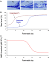
Cochlear postnatal development and functional maturation in mice. (A) Postnatal development of the cochlea. Arrows indicate that the cochlear tunnel starts to open at postnatal day 5 (P5) and fully opens at P10. (B) Postnatal developments of endocochlear potential (EP) and [K+]. Modified from Hibino et al. (2004). (C) Hearing maturation in mice. ABR thresholds dramatically drop at P11–16 and reach normal levels around P20. Modified from Liang et al. (2012).
Figure 3
Figure 3
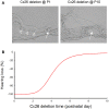
Cochlear developmental disorders and hearing loss induced by deletion of Cx26 in the cochlea at different postnatal times. (A) Cochlear development after deletion of Cx26 at P1 and P10. White arrows indicate that the cochlear tunnel is filled when Cx26 was deleted at P1 but developed normally when Cx26 was deleted at P10. Empty triangles indicate lack of the under-tectorial-membrane space; the tectorial membrane is attached to the inner sulcus cells following deletion of Cx26 at P1. (B) Hearing loss following deletion of Cx26 at different postnatal times. The ABR thresholds were measured at P30 and were normalized to that in WT mice. Corresponding to cochlear developmental disorders, deletion of Cx26 before P5 can induce severe hearing loss. However, hearing remains normal in young mice following deletion of Cx26 after P6. Modified from Chen et al. (2014).
Figure 4
Figure 4
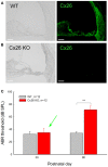
Normal hearing in young Cx26 KO mice after deletion of Cx26 after birth. (A,B) Immunofluorescent staining for Cx26 in the cochlea in WT and Cx26 KO mice at P30 following deletion of Cx26 at P10. No positive labeling is visible in the Cx26 KO mouse. (C) ABR thresholds of WT and Cx26 KO mice were measured at P30 and P90 following deletion of Cx26 at P10. A green arrow indicates that the ABR threshold in Cx26 KO mice at P30 remained normal, even though the expression of Cx26 in the cochlea was already deleted [see (B)]. At P90, Cx26 KO mice had a significant increase in ABR threshold, displaying hearing loss. **, P < 0.001, t-test. Scale bars: 50 μm. Modified from Zhu et al. (2015).

References

    1. Ahmad S., Tang W., Chang Q., Qu Y., Hibshman J., Li Y., et al. (2007). Restoration of connexin26 protein level in the cochlea completely rescues hearing in a mouse model of human connexin30-linked deafness. Proc. Natl. Acad. Sci. U.S.A. 104, 1337–1341.10.1073/pnas.0606855104 - DOI - PMC - PubMed
    1. Ashmore J. (2008). Cochlear outer hair cell motility. Physiol. Rev. 88, 173–210.10.1152/physrev.00044.2006 - DOI - PubMed
    1. Baranova A., Ivanov D., Petrash N., Pestova A., Skoblov M., Kelmanson I., et al. (2004). The mammalian pannexin family is homologous to the invertebrate innexin gap junction proteins. Genomics 83, 706–716.10.1016/j.ygeno.2003.09.025 - DOI - PubMed
    1. Beltramello M., Bicego M., Piazza V., Ciubotaru C. D., Mammano F., D’Andrea P. (2003). Permeability and gating properties of human connexins 26 and 30 expressed in HeLa cells. Biochem. Biophys. Res. Commun. 305, 1024–1033.10.1016/S0006-291X(03)00868-4 - DOI - PubMed
    1. Beltramello M., Piazza V., Bukauskas F. F., Pozzan T., Mammano F. (2005). Impaired permeability to Ins(1,4,5)P3 in a mutant connexin underlies recessive hereditary deafness. Nat. Cell Biol. 7, 63–69.10.1038/ncb1205 - DOI - PubMed