

Input-output relation and energy efficiency in the neuron with different spike threshold dynamics
- PMID: 26074810
- PMCID: PMC4444831
- DOI: 10.3389/fncom.2015.00062
Input-output relation and energy efficiency in the neuron with different spike threshold dynamics
Abstract
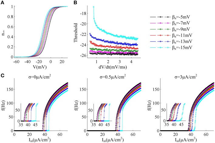
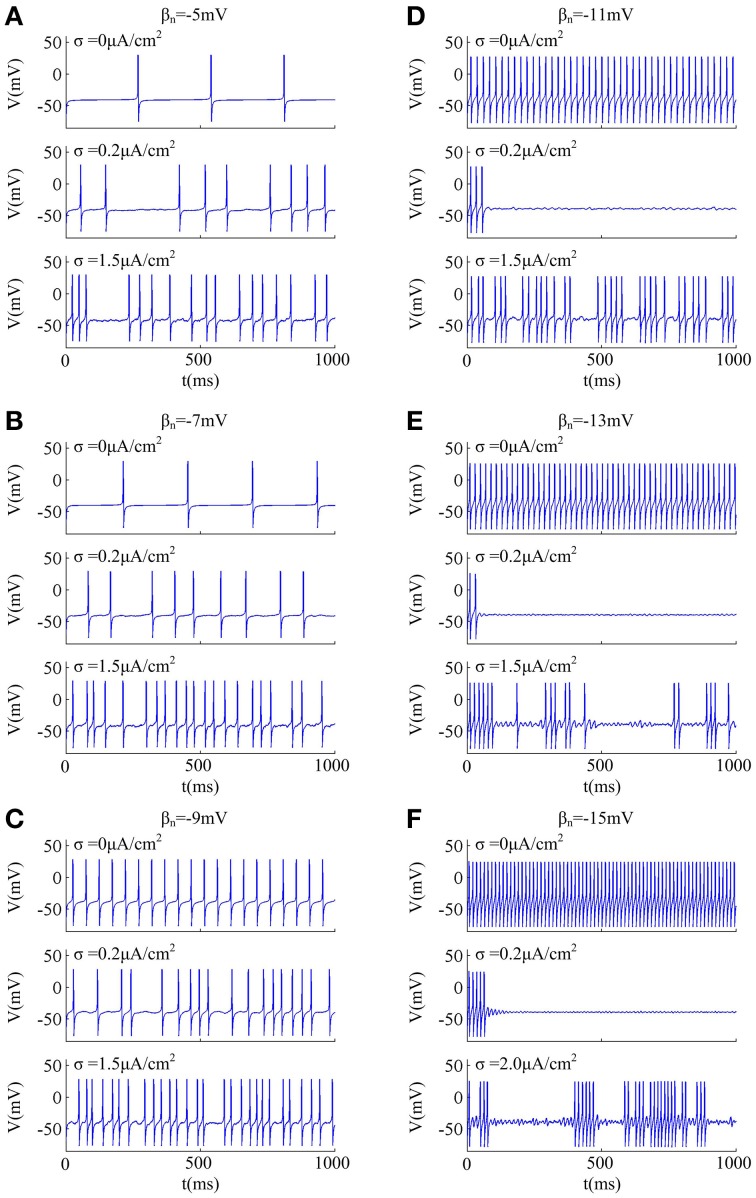
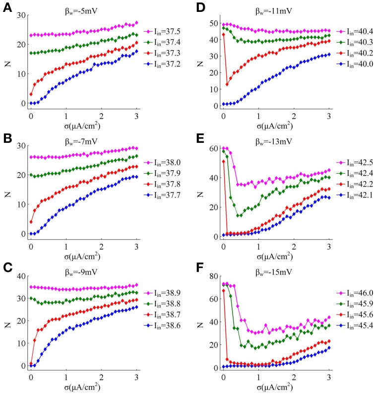
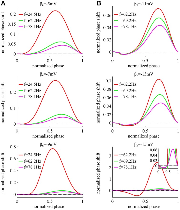
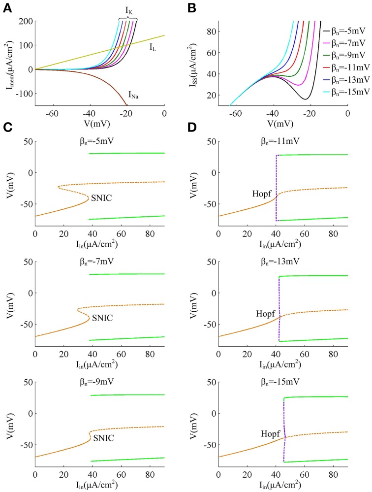
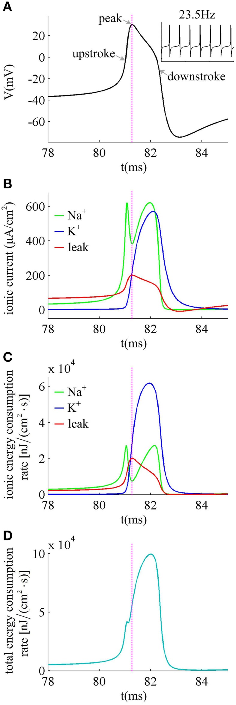
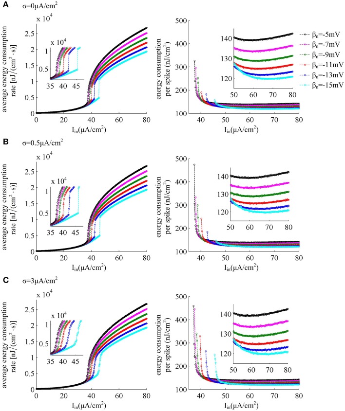
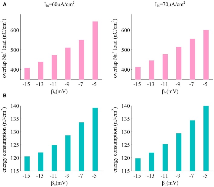
Neuron encodes and transmits information through generating sequences of output spikes, which is a high energy-consuming process. The spike is initiated when membrane depolarization reaches a threshold voltage. In many neurons, threshold is dynamic and depends on the rate of membrane depolarization (dV/dt) preceding a spike. Identifying the metabolic energy involved in neural coding and their relationship to threshold dynamic is critical to understanding neuronal function and evolution. Here, we use a modified Morris-Lecar model to investigate neuronal input-output property and energy efficiency associated with different spike threshold dynamics. We find that the neurons with dynamic threshold sensitive to dV/dt generate discontinuous frequency-current curve and type II phase response curve (PRC) through Hopf bifurcation, and weak noise could prohibit spiking when bifurcation just occurs. The threshold that is insensitive to dV/dt, instead, results in a continuous frequency-current curve, a type I PRC and a saddle-node on invariant circle bifurcation, and simultaneously weak noise cannot inhibit spiking. It is also shown that the bifurcation, frequency-current curve and PRC type associated with different threshold dynamics arise from the distinct subthreshold interactions of membrane currents. Further, we observe that the energy consumption of the neuron is related to its firing characteristics. The depolarization of spike threshold improves neuronal energy efficiency by reducing the overlap of Na(+) and K(+) currents during an action potential. The high energy efficiency is achieved at more depolarized spike threshold and high stimulus current. These results provide a fundamental biophysical connection that links spike threshold dynamics, input-output relation, energetics and spike initiation, which could contribute to uncover neural encoding mechanism.
Keywords: biophysical connection; energy efficiency; input-output relation; spike initiation; spike threshold dynamic.
Figures
Similar articles
-
Biophysical Insights into How Spike Threshold Depends on the Rate of Membrane Potential Depolarization in Type I and Type II Neurons.PLoS One. 2015 Jun 17;10(6):e0130250. doi: 10.1371/journal.pone.0130250. eCollection 2015. PLoS One. 2015. PMID: 26083350 Free PMC article.
-
How adaptation currents change threshold, gain, and variability of neuronal spiking.J Neurophysiol. 2014 Mar;111(5):939-53. doi: 10.1152/jn.00586.2013. Epub 2013 Oct 30. J Neurophysiol. 2014. PMID: 24174646
-
Auditory nerve spike generator modeled as a variable attenuator based on a saddle node on invariant circle bifurcation.PLoS One. 2012;7(9):e45326. doi: 10.1371/journal.pone.0045326. Epub 2012 Sep 18. PLoS One. 2012. PMID: 23028935 Free PMC article.
-
Biophysical basis for three distinct dynamical mechanisms of action potential initiation.PLoS Comput Biol. 2008 Oct;4(10):e1000198. doi: 10.1371/journal.pcbi.1000198. Epub 2008 Oct 10. PLoS Comput Biol. 2008. PMID: 18846205 Free PMC article.
-
The century-old picture of a nerve spike is wrong: filaments fire, before membrane.Commun Integr Biol. 2022 May 10;15(1):115-120. doi: 10.1080/19420889.2022.2071101. eCollection 2022. Commun Integr Biol. 2022. PMID: 35574158 Free PMC article. Review.
Cited by
-
Dendritic Properties Control Energy Efficiency of Action Potentials in Cortical Pyramidal Cells.Front Cell Neurosci. 2017 Sep 1;11:265. doi: 10.3389/fncel.2017.00265. eCollection 2017. Front Cell Neurosci. 2017. PMID: 28919852 Free PMC article.
-
Dynamical Mechanism of Hyperpolarization-Activated Non-specific Cation Current Induced Resonance and Spike-Timing Precision in a Neuronal Model.Front Cell Neurosci. 2018 Mar 8;12:62. doi: 10.3389/fncel.2018.00062. eCollection 2018. Front Cell Neurosci. 2018. PMID: 29568262 Free PMC article.
-
Monitoring time domain characteristics of Parkinson's disease using 3D memristive neuromorphic system.Front Comput Neurosci. 2023 Dec 15;17:1274575. doi: 10.3389/fncom.2023.1274575. eCollection 2023. Front Comput Neurosci. 2023. PMID: 38162516 Free PMC article.
-
Spiking Neural Network (SNN) With Memristor Synapses Having Non-linear Weight Update.Front Comput Neurosci. 2021 Mar 11;15:646125. doi: 10.3389/fncom.2021.646125. eCollection 2021. Front Comput Neurosci. 2021. PMID: 33776676 Free PMC article.
-
A multilayer-multiplexer network processing scheme based on the dendritic integration in a single neuron.AIMS Neurosci. 2022 Feb 28;9(1):76-113. doi: 10.3934/Neuroscience.2022006. eCollection 2022. AIMS Neurosci. 2022. PMID: 35434280 Free PMC article.
References
LinkOut - more resources
Full Text Sources
Other Literature Sources