. 2015:2015:649080.
doi: 10.1155/2015/649080. Epub 2015 May 5.

A Double-Switch Cell Fusion-Inducible Transgene Expression System for Neural Stem Cell-Based Antiglioma Gene Therapy

Affiliations

A Double-Switch Cell Fusion-Inducible Transgene Expression System for Neural Stem Cell-Based Antiglioma Gene Therapy

Yumei Luo et al. Stem Cells Int. 2015.

Abstract

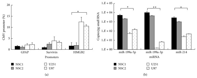
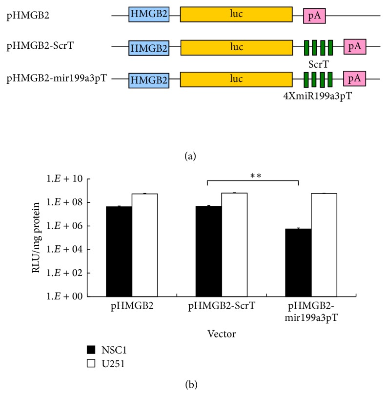
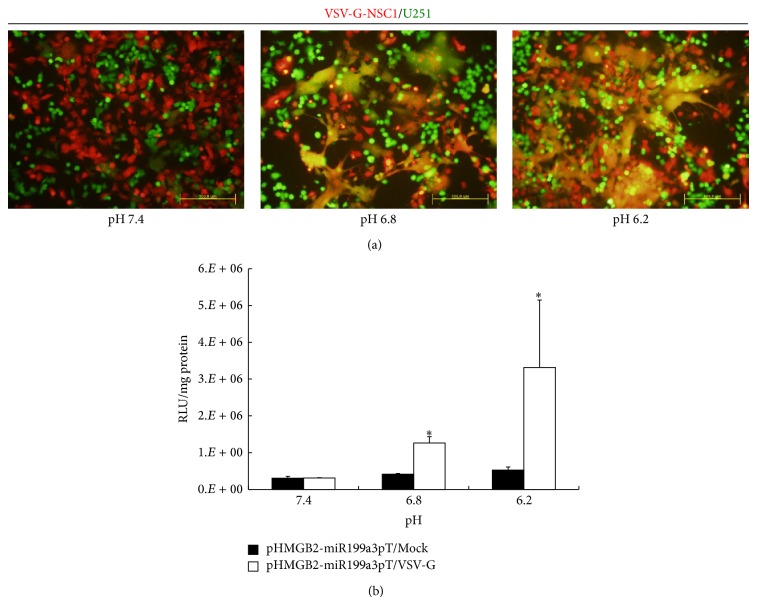
Recent progress in neural stem cell- (NSC-) based tumor-targeted gene therapy showed that NSC vectors expressing an artificially engineered viral fusogenic protein, VSV-G H162R, could cause tumor cell death specifically under acidic tumor microenvironment by syncytia formation; however, the killing efficiency still had much room to improve. In the view that coexpression of another antitumoral gene with VSV-G can augment the bystander effect, a synthetic regulatory system that triggers transgene expression in a cell fusion-inducible manner has been proposed. Here we have developed a double-switch cell fusion-inducible transgene expression system (DoFIT) to drive transgene expression upon VSV-G-mediated NSC-glioma cell fusion. In this binary system, transgene expression is coregulated by a glioma-specific promoter and targeting sequences of a microRNA (miR) that is highly expressed in NSCs but lowly expressed in glioma cells. Thus, transgene expression is "switched off" by the miR in NSC vectors, but after cell fusion with glioma cells, the miR is diluted and loses its suppressive effect. Meanwhile, in the syncytia, transgene expression is "switched on" by the glioma-specific promoter. Our in vitro and in vivo experimental data show that DoFIT successfully abolishes luciferase reporter gene expression in NSC vectors but activates it specifically after VSV-G-mediated NSC-glioma cell fusion.


Figures

Figure 1
Figure 1
Selection of glioma-specific promoter and NSC-specific miR. (a) Promoter activities of the lineage-specific promoter GFAP, tumor-specific promoter Survivin, and glioma-specific promoter HMGB2 compared to the strong universal promoter CMV in different NSC and glioma cell lines are quantified by luciferase assays. (b) Absolute expression levels of miR-199a/214 cluster members miR-199a-5p, miR-199a-3p, and miR-214 in different NSC and glioma cell lines are quantified by qPCR. miR copy numbers were calculated based on a standard curve generated using a synthetic LIN-4 RNA oligonucleotide. Error bars: s.d. * P < 0.05, ** P < 0.01.
Figure 2
Figure 2
Combinatory effect of optHRP and miR-199a-5p on transgene regulation. (a) Schematic representation of the combinatorial expression cassettes containing the HMGB2 promoter and miRNA target sequences. HMGB2, high mobility group box 2 gene promoter; luc, luciferase reporter gene; miR-199a-3p and scramble target sequences as detailed in Table 1 were inserted into 3′-UTR, respectively; pA, polyA signal. (b) Transgene expression levels of different expression cassettes within NSC1 and U251 cell lines are quantified by luciferase assays. Error bars: s.d. * P < 0.05, ** P < 0.01.
Figure 3
Figure 3
DoFIT mediates cell fusion-inducible transgene expression in vitro. (a) Low pH-dependent cell fusion between VSV-G-expressing NSC1 and U251 is examined by dual-color syncytia formation assays. (b) Transgene expression levels of cocultures between DoFIT-NSCs and U251 under different pH conditions are quantified by luciferase assays. Error bars: s.d. * P < 0.05, ** P < 0.01.
Figure 4
Figure 4
DoFIT mediates NSC-delivered glioma-specific transgene expression in vivo. (a) Bioluminescent images showing transgene expression in the glioma-bearing mice. U251 cells are inoculated into the right forebrains of the mice. After the gliomas develop, DoFIT-NSCs are inoculated into both the left and the right forebrains of the mice. Bioluminescent images are taken on day 1 and day 2 after NSC inoculation. The luminescent readings (photons/sec) of both left and right forebrains are indicated below the images. (b) Line graph showing trends of the averaged transgene expression levels in the glioma sites (right forebrain) and the normal sites (left forebrain) over day 1 and day 2 after NSC inoculation. Statistical significance of glioma sites versus normal sites at each time point was calculated by Student's t-test. Error bars: s.d. * P < 0.05.

References

    1. Aboody K. S., Brown A., Rainov N. G., et al. Neural stem cells display extensive tropism for pathology in adult brain: evidence from intracranial gliomas. Proceedings of the National Academy of Sciences of the United States of America. 2000;97(23):12846–12851. doi: 10.1073/pnas.97.23.12846. - DOI - PMC - PubMed
    1. Jurvansuu J., Zhao Y., Leung D. S. Y., et al. Transmembrane protein 18 enhances the tropism of neural stem cells for glioma cells. Cancer Research. 2008;68(12):4614–4622. doi: 10.1158/0008-5472.can-07-5291. - DOI - PubMed
    1. Zhao Y., Wang S. Human NT2 neural precursor-derived tumor-infiltrating cells as delivery vehicles for treatment of glioblastoma. Human Gene Therapy. 2010;21(6):683–694. doi: 10.1089/hum.2009.196. - DOI - PubMed
    1. Lee E. X., Lam D. H., Wu C., et al. Glioma gene therapy using induced pluripotent stem cell derived neural stem cells. Molecular Pharmaceutics. 2011;8(5):1515–1524. doi: 10.1021/mp200127u. - DOI - PubMed
    1. Chen C., Wang Y., Goh S. S. L., et al. Inhibition of neuronal nitric oxide synthase activity promotes migration of human-induced pluripotent stem cell-derived neural stem cells toward cancer cells. Journal of Neurochemistry. 2013;126(3):318–330. doi: 10.1111/jnc.12199. - DOI - PubMed

LinkOut - more resources