. 2015 Aug;11(8):611-7.
doi: 10.1038/nchembio.1858. Epub 2015 Jun 10.

Catalytic in vivo protein knockdown by small-molecule PROTACs

Affiliations

Catalytic in vivo protein knockdown by small-molecule PROTACs

Daniel P Bondeson et al. Nat Chem Biol. 2015 Aug.

Abstract

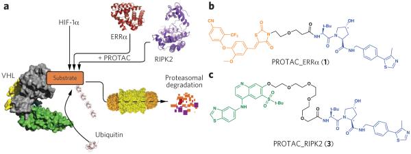
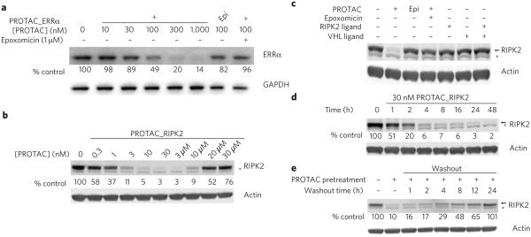
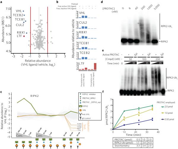
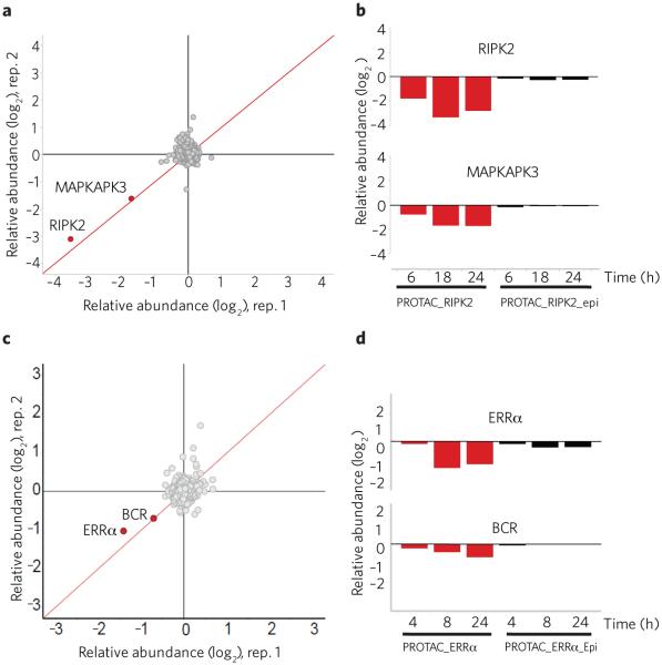
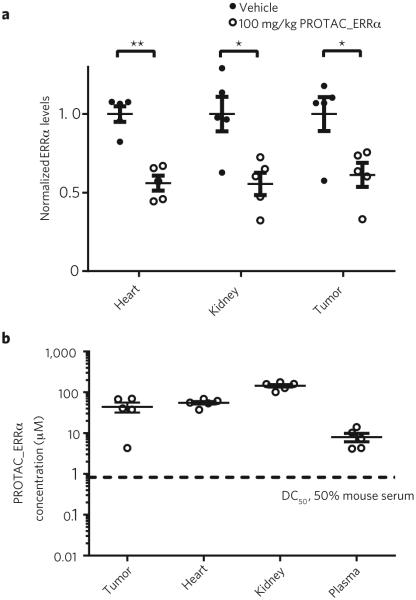
The current predominant therapeutic paradigm is based on maximizing drug-receptor occupancy to achieve clinical benefit. This strategy, however, generally requires excessive drug concentrations to ensure sufficient occupancy, often leading to adverse side effects. Here, we describe major improvements to the proteolysis targeting chimeras (PROTACs) method, a chemical knockdown strategy in which a heterobifunctional molecule recruits a specific protein target to an E3 ubiquitin ligase, resulting in the target's ubiquitination and degradation. These compounds behave catalytically in their ability to induce the ubiquitination of super-stoichiometric quantities of proteins, providing efficacy that is not limited by equilibrium occupancy. We present two PROTACs that are capable of specifically reducing protein levels by >90% at nanomolar concentrations. In addition, mouse studies indicate that they provide broad tissue distribution and knockdown of the targeted protein in tumor xenografts. Together, these data demonstrate a protein knockdown system combining many of the favorable properties of small-molecule agents with the potent protein knockdown of RNAi and CRISPR.


Figures

Figure 1
Figure 1
Proteolysis targeting chimeras (PROTACs). (a) Proposed model of PROTAC-induced degradation. Von Hippel–Lindau protein (VHL, gray) is an E3 ubiquitin ligase that, under normoxic conditions, functions with a cullin RING ligase (green and yellow) to degrade HIF1α. PROTACs recruit VHL to target proteins to induce their ubiquitination and subsequent proteasome-mediated downregulation. PROTACs were generated to two target proteins: the orphan nuclear receptor ERRα and the protein kinase RIPK2. (b) Structure of PROTAC_ERRα. The parent ERRα ligand is shown in orange and the modular VHL ligand in blue, with asterisks indicating stereocenter(s) whose inversion (in PROTAC_ERRα_epi) abolishes VHL binding. (c) Structure of PROTAC_RIPK2. The parent RIPK2 ligand is shown in green and the modular VHL ligand in blue, as in b.
Figure 2
Figure 2
PROTACs downregulate the protein levels of their respective targets. (a) PROTAC_ERRα dose-dependently downregulates ERRα protein levels. MCF7 cells were treated with either PROTAC_ERRα or PROTAC_ERRα_epi as indicated for 8 h before harvesting. Where indicated, cells were pretreated with 1 μM of the proteasome inhibitor epoxomicin for 1 h before the treatment. Target protein levels were subsequently detected by western blot analysis. Protein levels were normalized to loading and DMSO controls. Unless otherwise noted, all results in this work are representative of at least three independent experiments. (b) PROTAC_RIPK2 dose-dependently downregulates protein levels and demonstrates an amelioration of efficacy at higher concentrations (‘hook effect’) consistent with a ternary complex–mediated mechanism. THP-1 cells were treated with the indicated amounts of RIPK2_PROTAC for 16 h and then analyzed by western blotting. (c) Degradation by PROTACs is dependent on the proteasome and the presence of the linkage between both targeting ligands. THP-1 cells were treated with the indicated compounds (1 μM for epoxomicin, 30 nM for all others) for 16 h and analyzed by western blotting. (d) RIPK2 is rapidly degraded by PROTAC_RIPK2. THP-1 cells were treated with 30 nM PROTAC_RIPK2 for the indicated times and then analyzed by western blotting. (e) Downregulation by PROTACs is reversible. After a 4-h pretreatment with 30 nM PROTAC_RIPK2, the medium was replaced on THP-1 cells with fresh medium lacking PROTAC and the cells washed thoroughly to remove residual PROTAC. After the indicated times, the cells were analyzed by western blotting. In bd, * indicates a nonspecific band observed on western blots. For all panels, uncropped blots are shown in the corresponding panels of Supplementary Figure 6 (Supplementary Fig. 6ae).
Figure 3
Figure 3
PROTACs induce the catalytic ubiquitination of their target protein in a reconstituted E1-E2-VHL assay. (a) VHL ligand is highly selective for VHL and associated proteins. The VHL ligand was tethered to Sepharose beads and used to precipitate associated proteins. This was followed by washing of beads, elution of bound proteins and proteomic analysis of over 7,000 proteins. Detailed statistics are available in Online Methods. (b) VHL ligand binds selectively to VHL complex. Active (left three graphs) or inactive (right three graphs) VHL ligands were immobilized onto Sepharose beads and added to THP-1 cell lysates pretreated with active (experiments 1 and 4) or inactive (experiments 2 and 5) free VHL ligand or vehicle (experiments 3 and 6). Immobilized active VHL ligand selectively precipitated members of the VHL E3–dependent ligase complex (compare experiments 3 and 6). This effect was abrogated by prior treatment with free active VHL ligand (compare experiments 1 and 3) but not with inactive, epimeric VHL ligand (compare experiments 2 and 3). The only other protein significantly precipitated was lactotransferrin, which was associated and competed with free ligand when using both active and inactive VHL ligands. (c) PROTAC_RIPK2 mediates the co-immunoprecipitation of VHL and RIPK2. Cell lysates were immunoprecipitated with either IgG control (lanes 9, 10) or an anti-VHL antibody (lanes 1–8) in the presence of PROTAC_RIPK2 (lanes 1–3) or PROTAC_RIPK2_epi (lanes 4–6). The expression levels of over 7,000 proteins were quantified and normalized to the VHL precipitate without PROTAC present (lane 7). (d) PROTAC_RIPK2 mediates direct RIPK2 ubiquitination in vitro. RIPK2 was labeled by autophosphorylation and then incubated with the indicated concentrations of PROTAC and the reconstituted ubiquitination cascade (see Online Methods for more details). Samples were quenched 15 min after initiation of the reaction, and imaged by PAGE and autoradiography. RIPK2-Ubn is indicated. (e) Increasing the PROTAC concentration increases the rate of ubiquitination. Reactions were performed as in d with 50, 100 or 200 nM PROTAC_RIPK2 or PROTAC_RIPK2_epi, quenched at the indicated times and then analyzed by PAGE. (f) PROTACs are able to induce super-stoichiometric ubiquitination of RIPK2. Bands corresponding to ‘Modified RIPK2’ (RIPK2 that had received any number of ubiquitins) were excised, and the number of moles of RIPK2 from two parallel experiments was determined (see Online Methods). Abundance of modified RIPK2 (in pmol) is plotted against time for the three different reactions employing the indicated amounts of PROTAC_RIPK2.
Figure 4
Figure 4
PROTACs are highly specific for their respective target. (a) PROTAC_RIPK2 is highly selective for RIPK2 degradation. THP-1 cells were treated for 18 h with 30 nM PROTAC_RIPK2 in biological duplicate and protein levels quantified (see Online Methods). Data is plotted as fold change (log2) of replicate 1 versus replicate 2. The red diagonal line represents proteins whose changes in protein levels were reproducible between the two experiments. A total of 7,640 proteins were quantified. (b) THP-1 cells were treated for the indicated times with 30 nM of either PROTAC_RIPK2 or PROTAC_RIPK2_epi as in a, and the quantified levels of RIPK2 and MAPKAPK3 are shown. (c) MCF-7 cells were treated for 24 h with 500 nM PROTAC_ERRα in biological duplicate and protein levels quantified. Data is plotted as fold change (log2) of replicate 1 versus replicate 2. The red diagonal line represents proteins whose changes in protein levels were reproducible between the two experiments. A total of 7,576 proteins were quantified. (d) MCF-7 cells were treated for the indicated times with 500 nM PROTAC_ERRα or PROTAC_ERRα_epi as in c, and the quantified levels of ERRα and BCR are shown.
Figure 5
Figure 5
PROTAC_ERRα is efficacious in mice. (a) Mice (n = 5) were injected with either vehicle or 100 mg/kg PROTAC_ERRα (3 times per day, intraperitoneally). At ~5 h after the last injection, the mice were killed, and their tissues and tumors were collected and analyzed for ERRα expression by western blotting. Levels of ERRα were normalized to GAPDH levels and plotted, and are shown as mean ± s.e.m. **P <= 0.005, *P < 0.05 by two-tailed, unpaired Student’s t-test. (b) Tissues and plasma from a were analyzed for levels of PROTAC_ERRα by LC/MS. The dashed line represents the DC50 of PROTAC_ERRα when the in vitro degradation experiments were performed in 50% mouse serum. Each data point represents the levels of PROTAC_ERRα from a single mouse and tissue. Data are plotted and shown as mean ± s.e.m.

Comment in

References

    1. Adjei AA. What is the right dose? The elusive optimal biologic dose in phase I clinical trials. J. Clin. Oncol. 2006;24:4054–4055. - PubMed
    1. Soutschek J, et al. Therapeutic silencing of an endogenous gene by systemic administration of modified siRNAs. Nature. 2004;432:173–178. - PubMed
    1. Bumcrot D, Manoharan M, Koteliansky V, Sah DW. RNAi therapeutics: a potential new class of pharmaceutical drugs. Nat. Chem. Biol. 2006;2:711–719. - PMC - PubMed
    1. Tokatlian T, Segura T. siRNA applications in nanomedicine. Wiley Interdiscip. Rev. Nanomed. Nanobiotechnol. 2010;2:305–315. - PMC - PubMed
    1. Buckley DL, Crews CM. Small-molecule control of intracellular protein levels through modulation of the ubiquitin proteasome system. Angew. Chem. Int. Edn. Engl. 2014;53:2312–2330. - PMC - PubMed

Publication types

MeSH terms

Substances