. 2015 Jul 1;125(7):2759-71.
doi: 10.1172/JCI80369. Epub 2015 Jun 15.

PHD2/3-dependent hydroxylation tunes cardiac response to β-adrenergic stress via phospholamban

PHD2/3-dependent hydroxylation tunes cardiac response to β-adrenergic stress via phospholamban

Liang Xie et al. J Clin Invest. .

Abstract

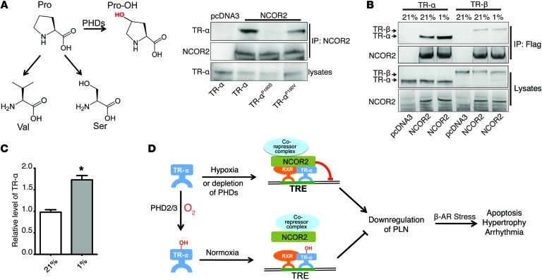
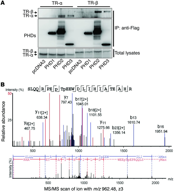
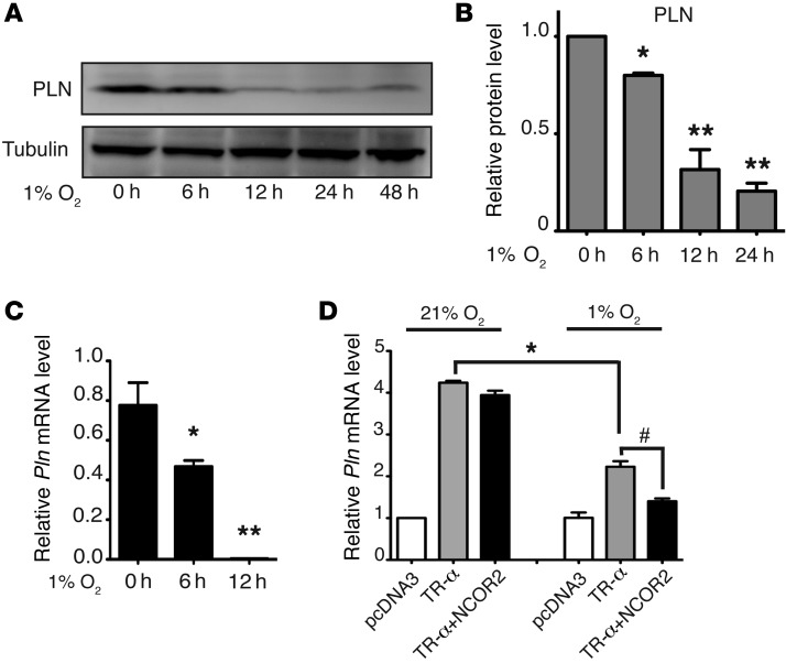
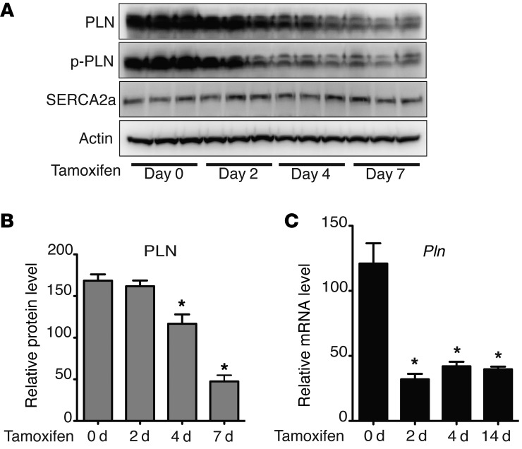
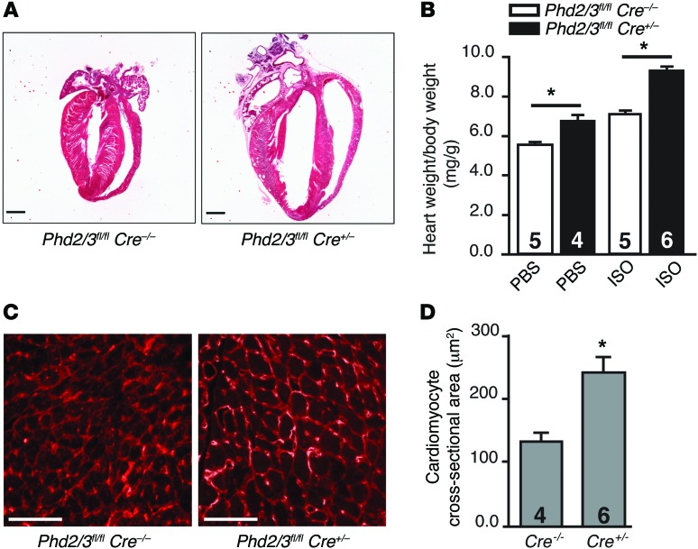
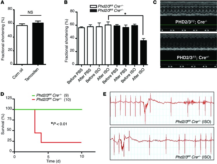
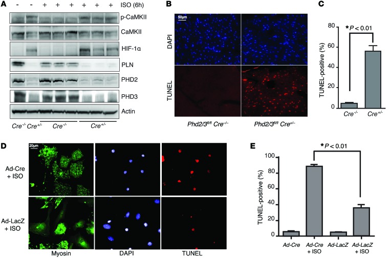
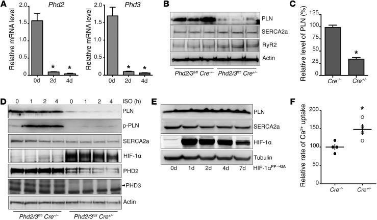
Ischemic heart disease is the leading cause of heart failure. Both clinical trials and experimental animal studies demonstrate that chronic hypoxia can induce contractile dysfunction even before substantial ventricular damage, implicating a direct role of oxygen in the regulation of cardiac contractile function. Prolyl hydroxylase domain (PHD) proteins are well recognized as oxygen sensors and mediate a wide variety of cellular events by hydroxylating a growing list of protein substrates. Both PHD2 and PHD3 are highly expressed in the heart, yet their functional roles in modulating contractile function remain incompletely understood. Here, we report that combined deletion of Phd2 and Phd3 dramatically decreased expression of phospholamban (PLN), resulted in sustained activation of calcium/calmodulin-activated kinase II (CaMKII), and sensitized mice to chronic β-adrenergic stress-induced myocardial injury. We have provided evidence that thyroid hormone receptor-α (TR-α), a transcriptional regulator of PLN, interacts with PHD2 and PHD3 and is hydroxylated at 2 proline residues. Inhibition of PHDs increased the interaction between TR-α and nuclear receptor corepressor 2 (NCOR2) and suppressed Pln transcription. Together, these observations provide mechanistic insight into how oxygen directly modulates cardiac contractility and suggest that cardiac function could be modulated therapeutically by tuning PHD enzymatic activity.


Figures

Figure 8
Figure 8. Hypoxia increases the interaction between TR-α and NCOR2.
(A) Replacement of the P160 residue of TR-α alters NCOR2-binding affinity. Molecular structures of proline, 4-hydroxy-proline, valine, and serine are shown. HEK293 cells were transfected with TR-α, TR-αP160S, or TR-αP160V, together with pcDNA3 or NCOR2, as indicated, followed by IP with NCOR2. Western blot analyses were then performed with the indicated antibodies. (B and C) Hypoxia potentiates the interaction between NCOR2 and TR-α. HEK293 cells cultured under 21% O2 conditions were transfected with constructs expressing TR-α or TR-β, together with pcDNA3 or NCOR2, as indicated, and were cultured under 21% or 1% O2 conditions for 8 hours. Flag-NCOR2 was IP, and Western blotting was performed with the indicated antibodies. Densitometric analysis of the IP TR-α is shown in C. n = 3, *P < 0.01, 2-tailed Student’s t test. (D) Schematic illustrating that PLN downregulation in the heart by hypoxia or PHD2 and PHD3 depletion contributes to myocardial injury induced by chronic β-AR stress. PHD2- and PHD3-mediated hydroxylation of TR-α blocks recruitment of the transcriptional repressor NCOR2 to the promoter region of Pln, resulting in transcription of Pln. Inhibition of TR-α hydroxylation, either through PHD2 and PHD3 depletion or through hypoxia, leads to increased recruitment of NCOR2 and suppression of Pln transcription. This decrease in PLN expression exacerbates cardiomyocyte apoptosis, cardiac hypertrophy, and arrhythmia induced by chronic β-AR stress.
Figure 7
Figure 7. TR-α is hydroxylated at proline residues P160 and P162.
(A) HL-1 cells were transfected with constructs expressing TR-α or TR-β, together with constructs expressing Flag-PHD1, -2, or -3, as indicated. Co-IP was performed with anti-Flag resin, and Western blot experiments were performed with the indicated antibodies. Both PHD2 and PHD3, but not PHD1, were able to pull down TR-α or TR-β. Representative blots from 3 experiments are shown. (B) An in vitro hydroxylation assay was performed with Flag–TR-α and PHD2/3. The protein band corresponding to TR-α was cut out for trypsin digestion. LC-MS/MS analysis was then performed. Tandem mass spectra of the precursor ion at m/z = 962.48 (Z = 3) for the human TR-α 153-176 sequence SLQQRPEP(+15.99)TP(+15.99)EEWDLIHIATEAHR are shown. The peak heights are the relative abundances of the corresponding fragment ions, with annotation of the identified matched N terminus–containing ions (b ions) shown in blue and C terminus–containing ions (y ions) shown in red. For clarity, only the major identified peaks are labeled (a complete table of fragment ions is presented in Supplemental Figure 3). Fragment ions at m/z = 952.66 (b8) and m/z = 1024.01 (y17)2+ represent characteristic ions that unambiguously identified P160–P162 double hydroxylation.
Figure 6
Figure 6. Hypoxia decreases PLN protein levels and suppresses Pln transcription.
Neonatal rat ventricular myocytes were treated with hypoxia (1% O2) as indicated. Cells were then harvested for protein or mRNA analysis. (A and B) Western blot analyses were performed with the indicated antibodies (A). Densitometric analysis from 3 experiments is shown in B. *P < 0.05; **P < 0.01, compared with 0 hours, 1-way ANOVA. (C) Real-time PCR was performed to analyze relative Pln mRNA levels. n = 3, *P < 0.05; **P < 0.01, compared with 0 hours, 1-way ANOVA. (D) HL-1 cardiomyocytes were transfected with a pGL3-Pln promoter (–156 to +64) luciferase construct, together with pcDNA3, or with a construct expressing TR-α or NCOR2, as indicated. Cells were also cotransfected with a construct expressing Renilla luciferase as the internal control. Twenty-four hours after transfection, cells were cultured under 21% or 1% O2 conditions for 24 hours. Cells were then harvested and luciferase activity determined with a luminometer. Relative luciferase activity was calculated from 3 separate experiments. *P < 0.05; #P < 0.01, 2-way ANOVA.
Figure 5
Figure 5. Dynamic regulation of PLN expression by depletion of PHD2/3.
Phd2 and Phd3 deletion induced a reduction of both PLN protein and Pln mRNA transcript levels. On days 0, 2, 4, 7, or 14 after tamoxifen infusion, Phd2/3fl/fl Cre+/– mouse hearts were harvested for Western blot (A) (each lane represents 1 heart) and real-time PCR (C) analyses. PLN protein levels were reduced in the absence of PHD2 and PHD3 compared with those in untreated mice. Densitometric analysis of PLN protein levels is shown in B. n = 4, *P < 0.05, compared with day 0, 1-way ANOVA. (C) Real-time PCR was performed to determine Pln mRNA levels in the hearts. n = 4, *P < 0.05, compared with day 0, 1-way ANOVA.
Figure 4
Figure 4. Depletion of PHD2/3 potentiates cardiac hypertrophy.
Phd2/3fl/flCre+/– and Phd2/3fl/fl Cre–/– mice were infused with tamoxifen for 5 consecutive days. Miniosmotic pumps were then implanted to chronically deliver PBS or ISO (20 mg/kg/d) for another 7 days. (A) Hearts were fixed and stained with H&E. Representative heart images of Phd2/3fl/fl Cre+/– and Phd2/3fl/fl Cre–/– mice treated with ISO are shown. The chamber size in Phd2- and Phd3-null hearts was dramatically increased compared with that in WT hearts. (B) The heart weight/body weight ratio is shown for the indicated group. n = 4–6 mice/group, *P < 0.05, 2-way ANOVA. (C and D) Representative cross sections of left ventricles from Phd2/3fl/fl Cre–/– and Phd2/3fl/fl Cre+/– mice treated with ISO and stained with WGA showed increased cardiomyocyte size in Phd2- and Phd3-null mice compared with that in WT mice (C). Cross-sectional areas of cardiomyocytes are shown in D. n = 4 or 6 mice/group, *P < 0.02, 2-tailed Student’s t test. Scale bars: 50 μm.
Figure 3
Figure 3. Depletion of PHD2/3 exaggerates myocardial injury induced by chronic treatment with ISO.
(A) Short-term deletion of Phd2 and Phd3 had no significant effect on cardiac function. ECG analyses of Phd2/3fl/fl Cre+/– mice were performed 2 weeks after day 1 of i.p. injection of tamoxifen or corn oil control (n = 5/group). NS, 2-tailed Student’s t test. (B and C) Deletion of PHD2 and PHD3 exacerbated cardiac dysfunction induced by ISO in female mice. Female Phd2/3fl/fl Cre+/– and Phd2/3fl/fl Cre–/– mice were i.p. injected with tamoxifen once daily for 5 days, followed by PBS or ISO infusion with miniosmotic pumps (20 mg/kg/d) for 7 days, and cardiac function was measured by ECG. Quantitative analysis of fractional shortening is shown in B (n = 5/group). *P < 0.05, 2-way ANOVA. Representative M-mode ECGs of Phd2/3fl/fl Cre+/– and Phd2/3fl/fl Cre–/– mice after 7 days’ treatment with ISO are shown in C. (D and E) Survival of male Phd2- and Phd3-null mice was significantly lower than that of WT mice. Male Phd2/3fl/fl Cre+/– and Phd2/3fl/fl Cre–/– mice were i.p. injected with tamoxifen once daily for 5 days, followed by infusion of PBS or ISO for 7 days. Kaplan-Meier survival curves for mice with the indicated genotypes are shown in D. *P < 0.01, log-rank test. Representative ECGs with severe cardiac arrhythmias observed in male Phd2/3fl/fl Cre+/– mice are shown in E.
Figure 2
Figure 2. Depletion of PHD2/3 leads to abnormal activation of CaMKII and increases cardiomyocyte apoptosis induced by ISO.
(A) Mice lacking PHD2 and PHD3 have elevated CaMKII activation compared with WT mice. Phd2/3fl/fl Cre+/– and Phd2/3fl/fl Cre–/– mice were i.p. injected with tamoxifen once daily for 5 days. Mice were then treated with ISO or PBS for 6 hours on day 7 after the first tamoxifen injection. Heart lysates were immunoblotted with the indicated antibodies. Each lane represents heart lysate from 1 mouse. (B and C) Deletion of PHD2 and PHD3 potentiates ISO-induced apoptosis in cardiomyocytes. Male Phd2/3fl/fl Cre+/– and Phd2/3fl/fl Cre–/– mice were i.p. injected with tamoxifen once daily for 5 days, followed by ISO infusion with miniosmotic pumps (20 mg/kg/d) for 2 days. Hearts were harvested and fixed for H&E or TUNEL staining (B). Graph in C shows quantitative analyses. n = 5, P < 0.01, 2-tailed Student’s t test. (D and E) Primary isolated cardiomyocytes deficient in PHD2 and PHD3 were also sensitized to ISO-induced apoptosis. Phd2/3fl/fl cardiomyocytes were isolated and infected with Ad-Cre or LacZ for 2 days and then treated with PBS or ISO for another 24 hours. Myosin and TUNEL staining was then performed. Representative images are shown in D. Quantitative analysis of TUNEL staining from 3 experiments is shown in E. *P < 0.01, 2-way ANOVA. Scale bars: 50 μm (B), 20 μm (D).
Figure 1
Figure 1. Depletion of PHD2/3 results in decreased PLN protein levels and increased SR Ca2+ uptake.
Phd2/3fl/flCre+/– mice were i.p. injected with tamoxifen once daily for 5 consecutive days. (A) Hearts were harvested on the indicated day after the first injection of tamoxifen, and real-time PCR was performed to determine the relative mRNA levels of Phd2 and Phd3. Phd2 and Phd3 mRNA levels were significantly decreased after 2 days of tamoxifen injection. n = 4, *P < 0.01, compared with day 0, 1-way ANOVA. (B and C) Seven days after the first injection of tamoxifen, mouse hearts were harvested, and Western blot analysis was performed. Densitometric analysis of PLN protein levels is shown in C. PLN, but not SERCA2a or RyR2, protein levels were significantly decreased. n = 6, *P < 0.01, 2-tailed Student’s t test. (D) Neonatal ventricular myocytes were isolated from Phd2/3fl/fl Cre+/– and Phd2/3fl/fl Cre–/– mice. After treatment with 4-OHT for 5 days, myocytes were treated with ISO for 0, 1, 2, or 4 hours. Western blot analysis was then performed. (E) After infection with adenovirus expressing normoxia-stable HIF-1α for 0, 1, 2, 4, and 7 days, neonatal rat ventricular myocytes were harvested, and Western blot analysis was performed. p-PLN, phosphorylated PLN. (F) SR Ca2+ uptake rates were measured using heart homogenates from Phd2/3fl/fl Cre+/– and Phd2/3fl/fl Cre–/– mice after treatment with tamoxifen for 7 days. The Ca2+ uptake rate was increased in mice lacking PHD2 and PHD3. n = 4, *P < 0.05, 2-tailed Student’s t test.

References

    1. Roger VL, et al. Heart disease and stroke statistics — 2012 update: a report from the American Heart Association. Circulation. 2012;125(1):e2–e220. doi: 10.1161/CIR.0b013e31823ac046. - DOI - PMC - PubMed
    1. Schomig A. Adrenergic mechanisms in myocardial infarction: cardiac and systemic catecholamine release. J Cardiovasc Pharmacol. 1988;12(suppl 1):S1–S7. - PubMed
    1. Desantiago J, et al. Arrhythmogenic effects of β2-adrenergic stimulation in the failing heart are attributable to enhanced sarcoplasmic reticulum Ca load. Circ Res. 2008;102(11):1389–1397. doi: 10.1161/CIRCRESAHA.107.169011. - DOI - PMC - PubMed
    1. Bristow MR. β-Adrenergic receptor blockade in chronic heart failure. Circulation. 2000;101(5):558–569. doi: 10.1161/01.CIR.101.5.558. - DOI - PubMed
    1. Kushner FG, et al. 2009 Focused Updates: ACC/AHA Guidelines for the Management of Patients With ST-Elevation Myocardial Infarction (updating the 2004 Guideline and 2007 Focused Update) and ACC/AHA/SCAI Guidelines on Percutaneous Coronary Intervention (updating the 2005 Guideline and 2007 Focused Update): a report of the American College of Cardiology Foundation/American Heart Association Task Force on Practice Guidelines. Circulation. 2009;120(22):2271–2306. doi: 10.1161/CIRCULATIONAHA.109.192663. - DOI - PubMed

Publication types

MeSH terms

Substances