. 2015 Jun 15;10(6):e0128945.
doi: 10.1371/journal.pone.0128945. eCollection 2015.

Redundancy between Cysteine Cathepsins in Murine Experimental Autoimmune Encephalomyelitis

Affiliations

Redundancy between Cysteine Cathepsins in Murine Experimental Autoimmune Encephalomyelitis

Euan Ramsay Orr Allan et al. PLoS One. .

Abstract

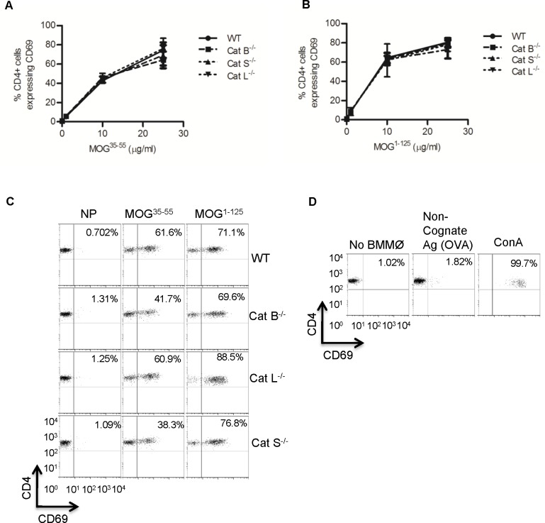
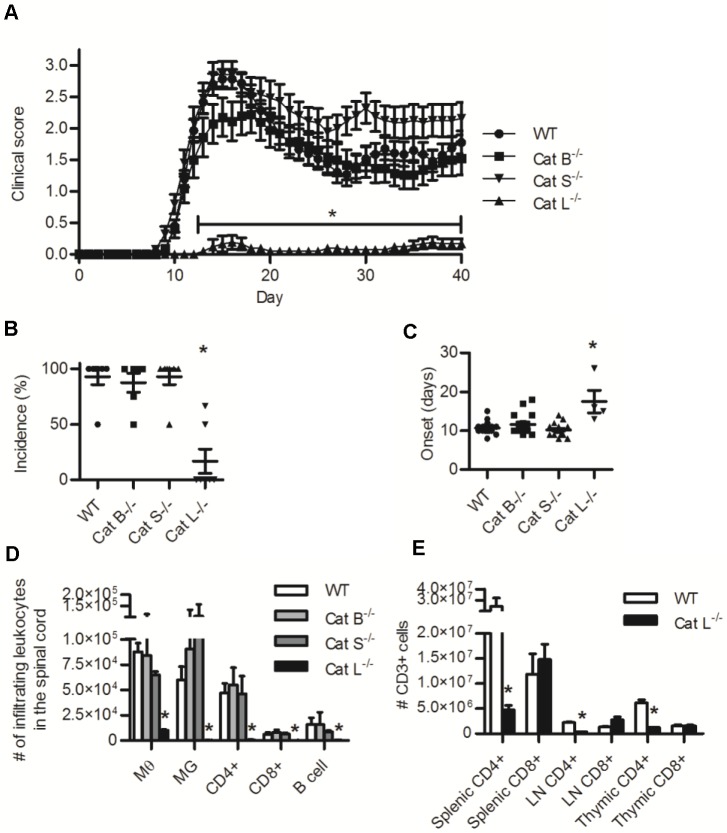
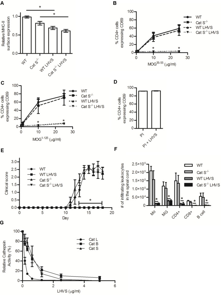
The cysteine cathepsins B, S, and L are functionally linked to antigen processing, and hence to autoimmune disorders such as multiple sclerosis. Stemming from several studies that demonstrate that mice can be protected from experimental autoimmune encephalomyelitis (EAE) through the pharmacologic inhibition of cysteine cathepsins, it has been suggested that targeting these enzymes in multiple sclerosis may be of therapeutic benefit. Utilizing mice deficient in cysteine cathepsins both individually and in combination, we found that the myelin-associated antigen myelin oligodendrocyte glycoprotein (MOG) was efficiently processed and presented by macrophages to CD4+ T cells in the individual absence of cathepsin B, S or L. Similarly, mice deficient in cathepsin B or S were susceptible to MOG-induced EAE and displayed clinical progression and immune infiltration into the CNS, similar to their wild-type counterparts. Owing to a previously described CD4+ T cell deficiency in mice deficient in cathepsin L, such mice were protected from EAE. When multiple cysteine cathepsins were simultaneously inhibited via genetic deletion of both cathepsins B and S, or by a cathepsin inhibitor (LHVS), MHC-II surface expression, MOG antigen presentation and EAE were attenuated or prevented. This study demonstrates the functional redundancy between cathepsin B, S and L in EAE, and suggests that the inhibition of multiple cysteine cathepsins may be needed to modulate autoimmune disorders such as multiple sclerosis.


Conflict of interest statement

Competing Interests: The authors have declared that no competing interests exist.

Figures

Fig 1
Fig 1. BMMØs derived from WT mice and mice deficient in cathepsin B, S, or L process and present MHC-II-restricted MOG antigens equivalently.
WT, cathepsin B (Cat B-/-), cathepsin S (Cat S-/-), cathepsin L (Cat L-/-) deficient BMMØ were incubated for 6 h with (A) MOG35-55 peptide (0, 1, 10, 25 μg/ml) or (B) MOG1-125 (0, 1, 10, 25 μg/ml). Activation of MOG35-55-specific 2D2 CD4+ T cells was determined by surface expression of CD69 after 16 h exposure to the BMMØs. Data represent 4 independent experiments. Data presented as mean +/- SEM (ANOVA, p<0.05); significant differences from internal WT controls are denoted by asterisks (*). (C,D) Representative flow cytometry plots of samples containing 25 μg/ml MOG35-55 and MOG1-125, and control samples that contain no BMMØs, a non-cognate antigen (ovalbumin, OVA) and the T cell mitogen concanavalin A (ConA 5 μg/ml).
Fig 2
Fig 2. Mice deficient in cathepsin B or S are susceptible to MOG35-55 induced EAE while mice deficient in cathepsin L are protected from EAE and have a systemic deficiency of CD4+ T cells.
Induction of EAE was attempted in WT, cathepsin B (Cat B-/-), cathepsin S (Cat S-/-), and cathepsin L (Cat L-/-) deficient mice by inoculation of 50μg MOG35-55 in CFA (day 0) and 300 ng pertussis toxin (day 0 and 2). (A) Clinical scoring of EAE severity over 40 days post inoculation (N = 20–21 mice). (B) Clinical incidence (percentage of mice that exceeded a clinical score of 0). (C) Number of days to clinical onset (time between inoculation and a clinical score of at least 0.5). (D) Absolute numbers of macrophages (MØ, CD11b+/CD45 high), microglia (MG, CD11b+/CD45 low), CD4+ T cells (CD4+/CD3+), CD8+ T cells (CD8+/CD3+) and B cells (B220+/CD45+) isolated from the spinal cord (via a discontinuous percoll gradient) of mice 15 days following inoculation with MOG35-55 (n = 3–4). (E) The abundance of CD3+/CD4+ and CD3+/CD8+ cells in the spleen, lymph nodes, and thymus from WT and cathepsin L (Cat L-/-) deficient mice. Data presented as mean+/- SEM; significant differences (Clinical data, Kruskal-Wallis of daily clinical scores and total area under the curve; ANOVA or unpaired students t-test, p<0.05) from the WT internal control are denoted by asterisks (*).
Fig 3
Fig 3. LHVS treatment reduces MHC-II surface expression and MHC-II restricted presentation efficiencies of MOG antigens in BMMØ, and protects from EAE in WT and cathepsin S deficient mice.
(A-C) WT and cathepsin S (Cat S-/-) deficient BMMØ were treated with LHVS (5μg/ml) or DMSO (carrier) for 18 h before assessment of (A) MHC-II surface expression (I-Ab quantified by MFI) or their ability to activate MOG35-55-specific CD4+ T cells following incubation with (B) MOG35-55 peptide (0, 1, 10, 25 μg/ml), (C) MOG1-125 (0, 1, 10, 25 μg/ml), or (D) PMA (50 ng/ml) and ionomysin (1 μg/ml) (PI). Activation of MOG35-55-specific 2D2 CD4+ T cells was determined by surface expression of CD69 after 16 h incubation with the BMMØ (n = 4). (E) Clinical disease course of WT and Cat S-/- deficient mice after induction of EAE with 50μg MOG35-55 in CFA (day 0) and 300 ng pertussis toxin (day 0 and 2) with daily injection of 25 mg/kg LHVS or DMSO (carrier) (day 0–15) (n = 6). (F) Absolute numbers of macrophages (MØ, CD11b+/CD45 high), microglia (MG, CD11b+/CD45 low), CD4+ T cells (CD4+/CD3+), CD8+ T cells (CD8+/CD3+), and B cells (B220+/CD45+) isolated from the spinal cord (via a discontinuous percoll gradient) of mice 18 days following inoculation with MOG35-55 (n = 4). (G) The relative enzymatic activity of recombinant cathepsins B, S, and L in the presence of increasing concentrations of LHVS (n = 3). Relative activities were determined by the rate of increase in fluorescence of cathepsin-selective fluorogenic substrates at 37°C, pH 5.5, and expressed relative to uninhibited samples. Data presented as mean+/- SEM; significant differences (Clinical data, Kruskal-Wallis; ANOVA, p<0.05) from the WT internal control are denoted by asterisks (*).
Fig 4
Fig 4. BMMØ deficient in both cathepsins B and S have reduced MHC-II surface expression and presentation of MOG antigens, and mice deficient in both cathepsins B and S are protected from EAE.
(A-C) WT and cathepsin B and S (Cat B-/-S-/-) deficient BMMØ were (A) examined for their MHC-II surface expression (I-Ab, quantified by MFI), or their ability to activate MOG35-55-specific CD4+ T cells following incubation with (B) MOG35-55 peptide (0, 10, 25 μg/ml) or (C) MOG1-125 (0, 10, 25 μg/ml). Activation of MOG35-55-specific 2D2 CD4+ T cells was determined by surface expression of CD69 after 16 h incubation with the BMMØ (n = 4). (D) Clinical disease course and (E) number of days to clinical onset (score of at least 0.5) of WT and Cat B-/-S-/- deficient mice after active induction of EAE with 50μg MOG35-55 in CFA (day 0) and 300 ng pertussis toxin (day 0 and 2), (N = 8). (F) The number of macrophages (MØ, CD11b+/CD45 high), microglia (MG, CD11b+/CD45 low), CD4+ T cells (CD4+/CD3+), and CD8+ T cells (CD8+/CD3+) isolated from the spinal cord (via a discontinuous percoll gradient) of mice 18 days following inoculation with MOG35-55 (n = 3). Data presented as mean+/- SEM; significant differences (Clinical data, Mann-Whitney Test; unpaired students t-testp<0.05) from the WT internal control are denoted by asterisks (*).

References

    1. Vasiljeva O, Reinheckel T, Peters C, Turk D, Turk V, Turk B. Emerging roles of cysteine cathepsins in disease and their potential as drug targets. Curr Pharm Des. 2007;13(4):387–403. Epub 2007/02/22. . - PubMed
    1. Haves-Zburof D, Paperna T, Gour-Lavie A, Mandel I, Glass-Marmor L, Miller A. Cathepsins and their endogenous inhibitors cystatins: expression and modulation in multiple sclerosis. J Cell Mol Med. 2011;15(11):2421–9. Epub 2010/12/15. 10.1111/j.1582-4934.2010.01229.x . - DOI - PMC - PubMed
    1. Stoeckle C, Quecke P, Ruckrich T, Burster T, Reich M, Weber E, et al. Cathepsin S dominates autoantigen processing in human thymic dendritic cells. J Autoimmun. 2012;38(4):332–43. Epub 2012/03/20. 10.1016/j.jaut.2012.02.003S0896-8411(12)00026-1 [pii]. . - DOI - PubMed
    1. Beers C, Honey K, Fink S, Forbush K, Rudensky A. Differential regulation of cathepsin S and cathepsin L in interferon gamma-treated macrophages. J Exp Med. 2003;197(2):169–79. Epub 2003/01/23. - PMC - PubMed
    1. Honey K, Duff M, Beers C, Brissette WH, Elliott EA, Peters C, et al. Cathepsin S regulates the expression of cathepsin L and the turnover of gamma-interferon-inducible lysosomal thiol reductase in B lymphocytes. J Biol Chem. 2001;276(25):22573–8. Epub 2001/04/18. 10.1074/jbc.M101851200M101851200 [pii]. . - DOI - PubMed

Publication types

MeSH terms

LinkOut - more resources