. 2015 Jun 15;11(6):e1004954.
doi: 10.1371/journal.ppat.1004954. eCollection 2015 Jun.

Discordant Impact of HLA on Viral Replicative Capacity and Disease Progression in Pediatric and Adult HIV Infection

Affiliations

Discordant Impact of HLA on Viral Replicative Capacity and Disease Progression in Pediatric and Adult HIV Infection

Emily Adland et al. PLoS Pathog. .

Abstract

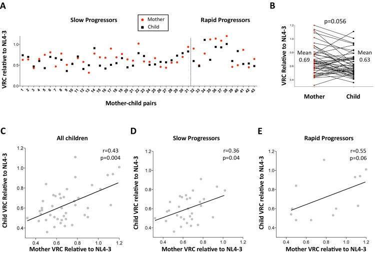
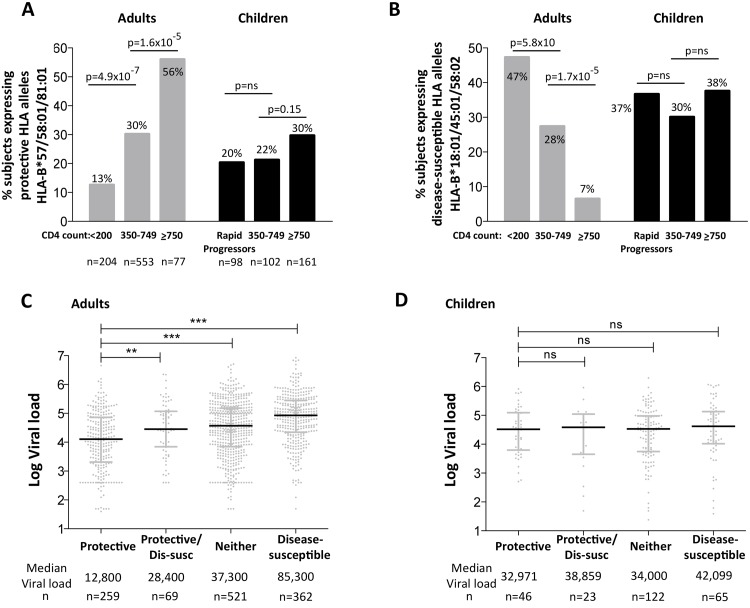
HLA class I polymorphism has a major influence on adult HIV disease progression. An important mechanism mediating this effect is the impact on viral replicative capacity (VRC) of the escape mutations selected in response to HLA-restricted CD8+ T-cell responses. Factors that contribute to slow progression in pediatric HIV infection are less well understood. We here investigate the relationship between VRC and disease progression in pediatric infection, and the effect of HLA on VRC and on disease outcome in adult and pediatric infection. Studying a South African cohort of >350 ART-naïve, HIV-infected children and their mothers, we first observed that pediatric disease progression is significantly correlated with VRC. As expected, VRCs in mother-child pairs were strongly correlated (p = 0.004). The impact of the protective HLA alleles, HLA-B*57, HLA-B*58:01 and HLA-B*81:01, resulted in significantly lower VRCs in adults (p<0.0001), but not in children. Similarly, in adults, but not in children, VRCs were significantly higher in subjects expressing the disease-susceptible alleles HLA-B*18:01/45:01/58:02 (p = 0.007). Irrespective of the subject, VRCs were strongly correlated with the number of Gag CD8+ T-cell escape mutants driven by HLA-B*57/58:01/81:01 present in each virus (p = 0.0002). In contrast to the impact of VRC common to progression in adults and children, the HLA effects on disease outcome, that are substantial in adults, are small and statistically insignificant in infected children. These data further highlight the important role that VRC plays both in adult and pediatric progression, and demonstrate that HLA-independent factors, yet to be fully defined, are predominantly responsible for pediatric non-progression.


Conflict of interest statement

The authors have declared that no competing interests exist.

Figures

Fig 1
Fig 1. Lower viral replicative capacity is associated with slow progression in children.
A. Replication capacities, normalized to NL4-3 comparator virus, were significantly lower in slow progressor children, median 0.61 (n = 55) compared to rapid progressor children, median 0.71 (n = 29) (p = 0.02). B. VRC correlation with decreasing absolute CD4 count in slow-progressor children at 5 years of age (n = 46). C. VRC correlation with decreasing absolute CD4 count in slow-progressor children at 5 years of age (n = 38). D. VRC correlation with decreasing absolute CD4 count in slow-progressor children at the age of assay date (n = 55).
Fig 2
Fig 2. Relationship between maternal VRC and pediatric VRC.
A. Maternal VRC (red circles) and child VRC (black squares) for each mother-child transmission pair. B. Comparison between VRC in mother and child: maternal mean VRC 0.69 versus VRC in the child 0.63; p = 0.056, paired t test. C. Correlation between VRC in mother and child for all children (n = 43, r = 0.43, p = 0.004). D. Correlation between VRC in mother and child for slow progressor children (n = 31, r = 0.36, p = 0.04). E. Correlation between VRC in mother and child for rapid progressor children (n = 12, r = 0.55, p = 0.06).
Fig 3
Fig 3. Relative contribution of HLA-B alleles in VRC in mothers and children.
A. VRC in mothers expressing protective HLA alleles (n = 16) and not (n = 30); and in children expressing protective HLA alleles (n = 22) and not (n = 54). B. VRC in mothers expressing disease-susceptible HLA alleles (n = 9) and not (n = 37); and in children expressing disease-susceptible alleles (n = 14) and not (n = 62). C. VRC in the child according to expression of protective HLA alleles in mother or child (+/+ n = 8;-/+ n = 8; +/- n = 9;-/- n = 18). D. VRC in the child according to expression of disease-susceptible alleles in the mother and/or child (+/+ n = 7;-/+ n = 7; +/- n = 6;-/- n = 23).
Fig 4
Fig 4. Impact of Gag mutations on VRC.
A-C: HLA-B*57/58:01/81:01 mutations. (A146X, I147X, A163X, S165X, E177X, Q182X, T186S, T242X, I247X and T310X). A. All subjects; B. Mothers; C. Children. D-F: Gag CD8+ T-cell mutations (listed in S1 Table). DS. All subjects. E. Mothers. F. Children.
Fig 5
Fig 5. Gag mutant number in adults with high VRC expressing disease-susceptible HLA.
A-B: HLA-B*57/58:01/81:01 mutations. A. Mothers. B; Children. C-D: Gag CD8+ T-cell mutations. C. Mothers. D. Children.
Fig 6
Fig 6. Impact of protective and disease-susceptible HLA alleles on disease progression in adults in children.
A. The frequency of ART-naïve adults and children stratified according to CD4 count and disease progression (see Methods) expressing one or more of the protective HLA alleles HLA-B*57/58:01/81:01. Adult study numbers: absolute CD4<200/mm3, n = 204; CD4 350–749/mm3, n = 553; CD4≥750/mm3 n = 77. Children study numbers: Rapid Progressors, n = 98; CD4 350–749/mm3, n = 102; CD4≥750/mm3, n = 161. B. As in panel A, but the frequency of subjects expressing one or more of the disease-susceptible HLA alleles HLA-B*18:01/45:01/58:02. C. Viral load in ART-naïve adults (total, n = 1,211) according to the presence of protective HLA alleles and/or disease-susceptible alleles. ** represents p<0.001, *** represents p<0.0001. Not shown on the figure are significant differences between viral loads in the disease-susceptible group and each of the other groups (in each case p<0.0001). D. As in panel C, but in ART-naïve children (total n = 256, age ≥5yrs).
Fig 7
Fig 7. Gag-specific CD8+ T-cell responses and autologous viral sequences in a rapid progressing child, P1.
A. Viral load and CD4% changes, CTL assays and viral sequencing timepoints. Closed red squares: viral load. Closed green squares: CD4%. Horizontal dotted lie: threshold for ART initiation (confirmed CD4<20%). Black closed circle: timepoint of maternal viral sequencing. Dbd: days before delivery. sdNVP: single dose nevirapine, given during labour and on the first day of life to the infant. D182: age of child at ART initiation. B. Gag-specific intracellular interferon-gamma staining assay undertaken when P1 was 10 days of age. C. Interferon-gamma elispot responses to Gag-33 and Gag-34 (two of 66 overlapping 18mers spanning the Gag protein), and on day 41 to the HLA-B*58:01-retricted Gag epitope TSTLQEQIAW contained within overlapping peptides Gag-33/4; and Gag-specific IFN-γ ICS data, showing responses to 66 pooled overlapping Gag peptides. D. Maternal viral sequence 31 days before delivery and autologous viral sequence in P1 showing lack of escape in all time points analyzed from day 1 to day 207, 25 days after ART initiation.
Fig 8
Fig 8. Rapid progression and lack of CTL escape in 4 additional rapid progressor expressing protective HLA-B*57/58:01/81:01.
A-D. Clinical data for subjects P2-P5 (as for P1 in Fig 7A). E. In child P5, elispot IFN-γ responses to 17–18mer Gag peptides Gag-40 (from day 4 of life) and Gag-25 (from day 79 of life) and Gag ICS responses to the pool of 66 Gag overlapping peptides. F. In child P5, recognition in IFN-γ elispot assays of autologous TL9 variants TPSDLNTML (Q182S) and TPSELNTML (D183E), compared to recognition of wildtype TL9. PBMC were tested at day 79 and day 141, with the pattern; data are shown from PBMC at day 141.

References

    1. Prendergast AJ, Klenerman P, Goulder PJ. The impact of differential antiviral immunity in children and adults. Nat Rev Immunol. 2012;12: 636–648. 10.1038/nri3277 - DOI - PubMed
    1. Pereyra F, Jia X, McLaren PJ, Telenti A, de Bakker PI, Walker BD, et al. The major genetic determinants of HIV-1 control affect HLA class I peptide presentation. Science 2010;330: 1551–1557. 10.1126/science.1195271 - DOI - PMC - PubMed
    1. Fellay J, Ge D, Shianna KV, Colombo S, Ledergerber B, Cirulli ET, et al. Common genetic variation and the control of HIV-1 in humans. PLoS Genet. 2009;5: e1000791 10.1371/journal.pgen.1000791 - DOI - PMC - PubMed
    1. Fellay J, Shianna KV, Ge D, Colombo S, Ledergerber B, Weale M, et al. A whole-genome association study of major determinants for host control of HIV-1. Science 2007;317: 944–947. - PMC - PubMed
    1. Goulder PJ, Walker BD. HIV and HLA class I: an evolving relationship. Immunity 2012;37: 426–440. 10.1016/j.immuni.2012.09.005 - DOI - PMC - PubMed

Publication types