. 2015 Jun 15;10(6):e0130389.
doi: 10.1371/journal.pone.0130389. eCollection 2015.

Derivation of Cinnamon Blocks Leukocyte Attachment by Interacting with Sialosides

Affiliations

Derivation of Cinnamon Blocks Leukocyte Attachment by Interacting with Sialosides

Wei-Ling Lin et al. PLoS One. .

Abstract

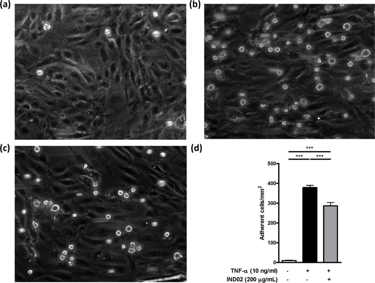
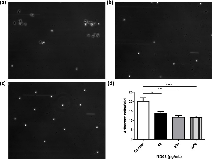
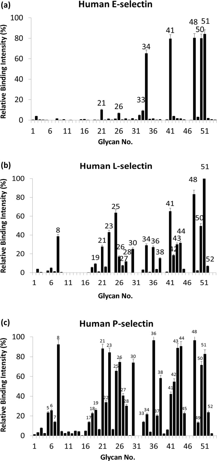
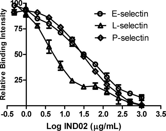
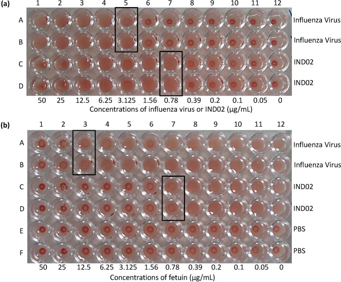
Molecules derived from cinnamon have demonstrated diverse pharmacological activities against infectious pathogens, diabetes and inflammatory diseases. This study aims to evaluate the effect of the cinnamon-derived molecule IND02 on the adhesion of leukocytes to host cells. The anti-inflammatory ability of IND02, a pentameric procyanidin type A polyphenol polymer isolated from cinnamon alcohol extract, was examined. Pretreatment with IND02 significantly reduced the attachment of THP-1 cells or neutrophils to TNF-α-activated HUVECs or E-selectin/ICAM-1, respectively. IND02 also reduced the binding of E-, L- and P-selectins with sialosides. Furthermore, IND02 could agglutinate human red blood cells (RBC), and the agglutination could be disrupted by sialylated glycoprotein. Our findings demonstrate that IND02, a cinnamon-derived compound, can interact with sialosides and block the binding of selectins and leukocytes with sialic acids.


Conflict of interest statement

Competing Interests: The authors have the following interests: Co-authors Ekambaranellore Prakash and Mohan Viswaraman are employed by Indus Biotech Private Limited. There are no patents, products in development or marketed products to declare. This does not alter the authors' adherence to all the PLOS ONE policies on sharing data and materials.

Figures

Fig 1
Fig 1. Monocytes adhesion assay.
(a) THP-1 monocytic cells were preincubated with PBS for 0.5 h at 37°C followed by perfusion into a flow chamber. The rolling (attachment) of THP-1 with HUVECs (without TNF-α activation) were recorded and captured. (b, c) THP-1 monocytic cells were preincubated with PBS (b) or IND02 (200 μg/mL) (c) for 0.5 h at 37°C followed by perfusion into flow chamber. The rolling (attachment) of THP-1 with TNF-α activated HUVECs were recorded and captured. (d) Control (without TNF-α activation): 9.86±1.81 cells/mm2; Control (TNF-α activated HUVEC): 378.5±11.5 cells/mm2; IND02 (200 μg/mL): 286.3±17.3 cells/mm2. The number of adherent cells was calculated in 10–15 random fields in a single experiment, and the statistical test were calculated based on 3–5 experiments using one-way ANOVA followed by post hoc test. The movie of (a)-(c) were showed in S1–S3 Movies. IND02 significantly reduced THP-1 attachment and rolling on TNF-α activated HUVECs. (***: P < 0.001).
Fig 2
Fig 2. Neutrophils attachment assay.
Neutrophils were preincubated with (a) PBS, (b) IND02 (1000 μg/mL), (c) IND02 (40 μg/mL), for 0.5 h at 37°C followed by perfusion into a flow chamber. The attachment of neutrophils with E-selectin/ICAM-1 coated slides were recorded and captured. (d) Control: 20.2±6.8 cells/field; IND02 (40 μg/mL): 13.7±3.6 cells/field; IND02 (200 μg/mL): 11.8±2.5 cells/field; IND02 (1000 μg/mL): 10.9±2.8 cells/field. The number of attached cells was calculated in 10–15 random fields in a single experiment, and the statistical test were calculated based on 3–5 experiments using one-way ANOVA followed by post hoc test. IND02 significantly reduced neutrophils attachment and rolling on E-selectin/ICAM-1 coated slides. (**: P < 0.01; ***: P < 0.001; ****: P < 0.0001).
Fig 3
Fig 3. Carbohydrate binding specificities of E-, L- and P-selectin.
Donor beads and biotin-PAA-sugars mixed with selectins were incubated at ambient temperature for 1 h (total 15 μL). The mixture of acceptor beads, mouse anti-human IgG antibody and rabbit anti-mouse IgG antibody was added to the reaction to a final volume of 25 μL. After 2 h of incubation, the binding signals were obtained using a PerkinElmer Envision instrument and the AlphaScreen program. The results were are indicated by relative intensities of the highest fluorescence signal (y-axis). The sugar identities are designated by numbers (x-axis) and shown in detail in S1 Table. Glycans which showed more than 30% relative intensity are indicated as number on each figure.
Fig 4
Fig 4. IND02 inhibited the binding of E-, L- and P-selectin to sialyl Lex.
E-, L- and P-selectin were incubated with biotin-PAA-sialyl Lex for 1 h in a 384 microtiter plate. Serially diluted IND02 (from 1000 μg/mL to 0.5 μg /mL) was added to each well, incubated at room temperature for 2 h and detected using a reader. Based on the relative binding intensity, the IC50 values of IND02 for E-, L- and P-selectin were 31.6, 5.0 and 28.2 μg/mL, respectively. The results were the average of six independent assays.
Fig 5
Fig 5
(a) Agglutination assays of the influenza virus and IND02. Two-fold serially diluted influenza A virus in row A and B (column 1 to 11, 50 μg/mL to 50 ng/mL) and two-fold serially diluted IND02 in row C and D (column 1 to 11, 50 μg/mL to 50 ng/mL) were prepared in the microtiter plate. Washed human RBCs (5% v/v) were added to the wells and incubated at 37°C for 1 h. The agglutination titers of influenza A virus and IND02 were 1:16 (3.125 μg/mL) and 1:64 (0.78125 μg/mL), respectively. (b) Inhibition assay. Two-fold serially diluted fetuin (column 1 to 11, 50 μg/mL to 0 ng/mL) were prepared. Influenza A virus (as control, row A and B, 10 μg/mL), IND02 (row C and D, 10 μg/mL), or PBS (row E and F) were added and incubated with fetuin at 37°C for 0.5 h. Washed human RBCs (5%) were then added to the reaction and incubated at 37°C for 1 h. The results showed that the lowest fetuin concentration that block human RBC agglutinations for the influenza virus and IND02 were 12.5 μg/mL and 0.78125 μg/mL, respectively.

References

    1. Kwon HK, Hwang JS, So JS, Lee CG, Sahoo A, Ryu JH, et al. Cinnamon extract induces tumor cell death through inhibition of NFkappaB and AP1. BMC Cancer. 2010;10: 392 10.1186/1471-2407-10-392 - DOI - PMC - PubMed
    1. Koppikar SJ, Choudhari AS, Suryavanshi SA, Kumari S, Chattopadhyay S, Kaul-Ghanekar R. Aqueous cinnamon extract (ACE-c) from the bark of Cinnamomum cassia causes apoptosis in human cervical cancer cell line (SiHa) through loss of mitochondrial membrane potential. BMC Cancer. 2010;10: 210 10.1186/1471-2407-10-210 - DOI - PMC - PubMed
    1. Lu J, Zhang K, Nam S, Anderson RA, Jove R, Wen W. Novel angiogenesis inhibitory activity in cinnamon extract blocks VEGFR2 kinase and downstream signaling. Carcinogenesis. 2010;31: 481–488. 10.1093/carcin/bgp292 - DOI - PMC - PubMed
    1. Kwon HK, Jeon WK, Hwang JS, Lee CG, So JS, Park JA, et al. Cinnamon extract suppresses tumor progression by modulating angiogenesis and the effector function of CD8+ T cells. Cancer Lett. 2009;278: 174–182. 10.1016/j.canlet.2009.01.015 - DOI - PubMed
    1. Shan B, Cai YZ, Brooks JD, Corke H. Antibacterial properties and major bioactive components of cinnamon stick (Cinnamomum burmannii): activity against foodborne pathogenic bacteria. J Agric Food Chem. 2007;55: 5484–5490. - PubMed

Publication types

MeSH terms

LinkOut - more resources