. 2015 Jun 16:5:10938.
doi: 10.1038/srep10938.

Anchor-based classification and type-C inhibitors for tyrosine kinases

Affiliations

Anchor-based classification and type-C inhibitors for tyrosine kinases

Kai-Cheng Hsu et al. Sci Rep. .

Abstract

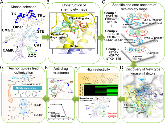
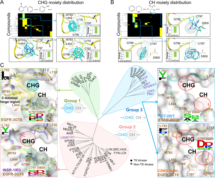
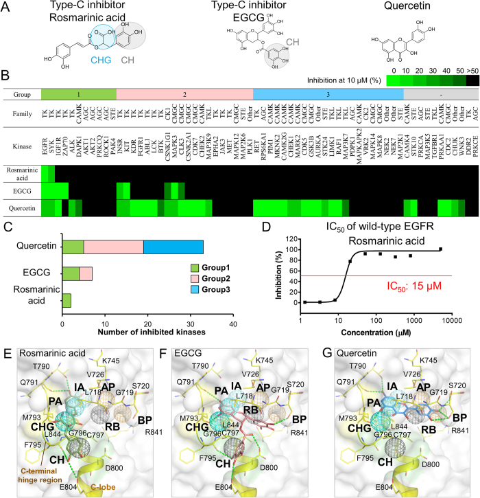
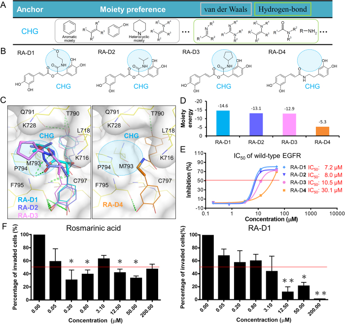
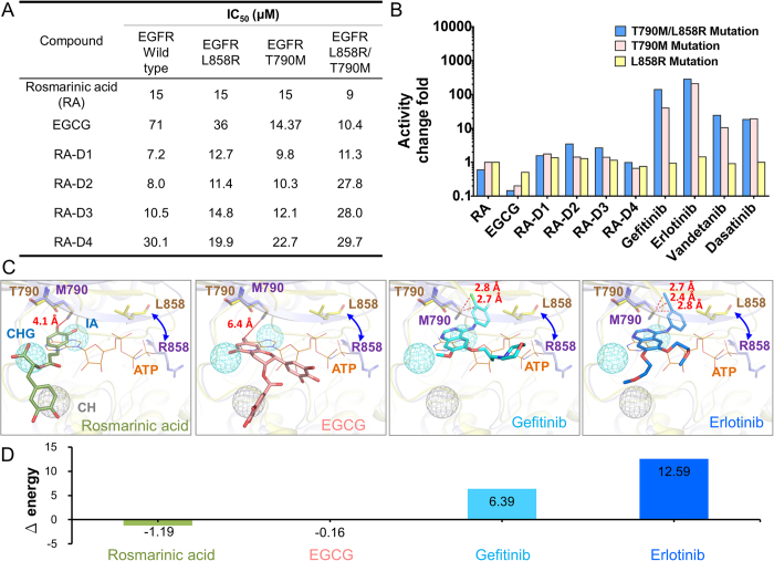
Tyrosine kinases regulate various biological processes and are drug targets for cancers. At present, the design of selective and anti-resistant inhibitors of kinases is an emergent task. Here, we inferred specific site-moiety maps containing two specific anchors to uncover a new binding pocket in the C-terminal hinge region by docking 4,680 kinase inhibitors into 51 protein kinases, and this finding provides an opportunity for the development of kinase inhibitors with high selectivity and anti-drug resistance. We present an anchor-based classification for tyrosine kinases and discover two type-C inhibitors, namely rosmarinic acid (RA) and EGCG, which occupy two and one specific anchors, respectively, by screening 118,759 natural compounds. Our profiling reveals that RA and EGCG selectively inhibit 3% (EGFR and SYK) and 14% of 64 kinases, respectively. According to the guide of our anchor model, we synthesized three RA derivatives with better potency. These type-C inhibitors are able to maintain activities for drug-resistant EGFR and decrease the invasion ability of breast cancer cells. Our results show that the type-C inhibitors occupying a new pocket are promising for cancer treatments due to their kinase selectivity and anti-drug resistance.


Figures

Figure 1
Figure 1. Overview of the quantification of binding specificity and discovery of type-C inhibitors for tyrosine kinases.
(A) Selection of 51 protein kinases. In total, 45 tyrosine kinases and 6 protein kinases from six other kinase families were chosen. (B) Construction of site-moiety maps for these 51 kinases using 4,680 known kinase inhibitors. (C) Identification of specific anchors for type-C kinase inhibitors. The cyan and red circles indicate two specific and five conserved anchors in the C-terminal hinge region and the ATP-binding site, respectively. Tyrosine kinases are classified into three groups based on the presence of these two specific anchors. (D) Comparison of type-C, type-I, and type-II inhibitors. The type-C inhibitors (rosmarinic acid, green) seize the specific anchors in the C-terminal hinge region, whereas type-I and type-II inhibitors occupy the ATP-binding site. (E) Two type-C inhibitors (rosmarinic acid and EGCG) and quercetin were tested against 64 protein kinases. (F) Enzyme-based drug resistance assays of rosmarinic acid on EGFR. (G) Lead optimization of type-C inhibitors guided by our anchor model.
Figure 2
Figure 2. Anchor-based classification of tyrosine kinases.
Interacting moiety profiles of (A) CHG and (B) CH anchors. An entry is colored yellow if the compound has a moiety that interacts with the anchor residues; otherwise, the entry is colored black. The frequently interacting functional groups and their average binding energy are shown near the profile. The moieties with a low binding energy can be used for the lead optimization process. (C) Classification tree and anchor residue patterns of three groups. The tree was constructed by considering the anchor presence and the anchor residue identities. Group 1 kinases contain CH and CHG anchors. The structures of EGFR and the other Group 1 kinases are shown as yellow stick and white line representations, respectively. Group 2 kinases lack the CHG anchor because of the long and rigid residue types (residue F/Y) at the 792 position near the CHG anchor. In Group 3 kinases, the short side chains of the residues at the positions 793 and 796 result in elimination of the CH and CHG anchors.
Figure 3
Figure 3. Selectivity of type-C inhibitors and quercetin.
(A) Structures of the compounds. Rosmarinic acid and EGCG are type-C inhibitors, which match two (CH and CHG) specific anchors and one (CH) specific anchor, respectively. In comparison, quercetin matches none of the specific anchors. (B) Results of compound profiling. The percentage of the remaining kinase activity is indicated from green (0%) to black (>50%). A compound is considered a potential inhibitor against kinases if the percentage of the remaining kinase activity is ≤50% at a compound concentration of 10 μM. (C) The target number of three compounds. Rosmarinic acid, EGCG, and quercetin inhibit 2 (3%), 9 (14%), and 39 (61%) of the 64 protein kinases, respectively. (D) IC50 value of rosmarinic acid for wild-type EGFR. Docked poses of the compounds (E) rosmarinic acid, (F) EGCG, and (G) quercetin in EGFR. Hydrogen-bonding interactions are represented as green dashes.
Figure 4
Figure 4. Specific anchor guides the synthesis of rosmarinic acid derivatives.
(A) Moiety preferences of CHG anchor. (B) Four rosmarinic acid derivatives, namely RA-D1, RA-D2, RA-D3, and RA-D4, were synthesized based on the moiety composition of the CHG anchor. (C)The moieties located at the CHG anchor are represented as sticks, and the other moieties are represented as lines. (D) Interaction energy between the substituted moieties and residues of the CHG anchor. (E) Dose-response curves and IC50 values of the derivatives. The first three derivatives have large substituted moieties that form van der Waals interactions with residues of the CHG anchor and consistently present better activity compared with rosmarinic acid. In comparison, RA-D4 lacks a moiety in the CHG anchor and shows poor inhibitory activity. (F) Effect of type-C inhibitors on invasion of MDA-MB-231 breast cancer cells. The invasion activity was tested using Corning BioCoat Matrigel Invasion Chambers. *, p < 0.05; **, p < 0.01, compared with the control group.
Figure 5
Figure 5. Type-C inhibitors against drug-resistant EGFR.
(A) IC50 values of type-C inhibitors against wild-type, T790M, L858R, and T790M/L858R mutant EGFR. (B) Fold changes in activity of rosmarinic acid, EGCG, gefitinib, erlotinib, vandetanib, and dasatinib in the presence of the drug-resistant mutations. (C) Binding modes of type-C inhibitors and kinase drugs in the T790M/L858R mutant EGFR. The kinase-drug complex structures and the mutant EGFR structure (PDB 3W2P) are superimposed on the structure of wild-type EGFR, including EGFR-gefitinib (PDB 2IVT) and EGFR-erlotinib (PDB 1M17) complex structures. (D) Energy difference of the compounds between the wild-type (T790) and mutant (M790) residues calculated by GEMDOCK. Type-C inhibitors maintain similar interaction energy for the mutant residue, whereas gefitinib and erlotinib lose interactions when the mutation occurs.

References

    1. Endicott J. A., Noble M. E. M. & Johnson L. N. The Structural Basis for Control of Eukaryotic Protein Kinases. Annu. Rev. Biochem. 81, 587–613 (2012). - PubMed
    1. Levitzki A. Tyrosine Kinase Inhibitors: Views of Selectivity, Sensitivity, and Clinical Performance. Annu. Rev. Pharmacol. Toxicol. 53, 161–185 (2013). - PubMed
    1. Krause D. S. & Van Etten R. A. Tyrosine kinases as targets for cancer therapy. N. Engl. J. Med. 353, 172–187 (2005). - PubMed
    1. Gocek E., Moulas A. N. & Studzinski G. P. Non-receptor protein tyrosine kinases signaling pathways in normal and cancer cells. Crit. Rev. Clin. Lab. Sci. 51, 125–137 (2014). - PubMed
    1. Liao J. J. L. Molecular recognition of protein kinase binding pockets for design of potent and selective kinase inhibitors. J. Med. Chem. 50, 409–424 (2007). - PubMed

Publication types

MeSH terms