. 2015 Jun 16:5:10948.
doi: 10.1038/srep10948.

Subgingival microbiome in patients with healthy and ailing dental implants

Affiliations

Subgingival microbiome in patients with healthy and ailing dental implants

Hui Zheng et al. Sci Rep. .

Abstract

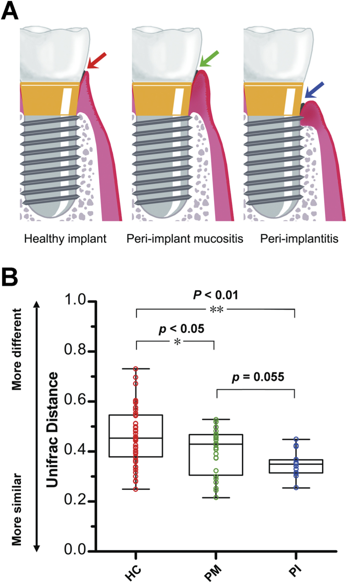
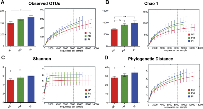
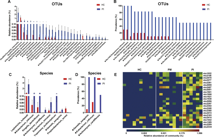
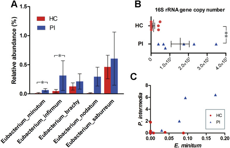
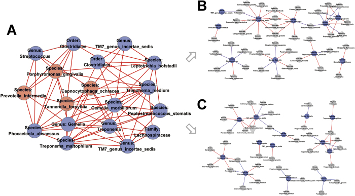
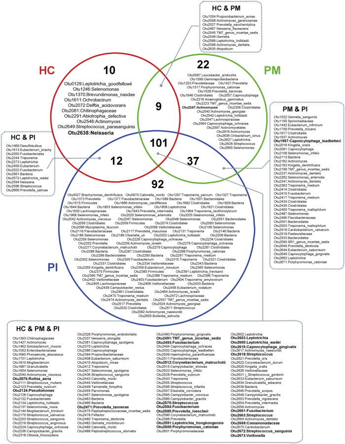
Dental implants are commonly used to replace missing teeth. However, the dysbiotic polymicrobial communities of peri-implant sites are responsible for peri-implant diseases, such as peri-implant mucositis and peri-implantitis. In this study, we analyzed the microbial characteristics of oral plaque from peri-implant pockets or sulci of healthy implants (n = 10), peri-implant mucositis (n = 8) and peri-implantitis (n = 6) sites using pyrosequencing of the 16S rRNA gene. An increase in microbial diversity was observed in subgingival sites of ailing implants, compared with healthy implants. Microbial co-occurrence analysis revealed that periodontal pathogens, such as Porphyromonas gingivalis, Tannerella forsythia, and Prevotella intermedia, were clustered into modules in the peri-implant mucositis network. Putative pathogens associated with peri-implantitis were present at a moderate relative abundance in peri-implant mucositis, suggesting that peri-implant mucositis an important early transitional phase during the development of peri-implantitis. Furthermore, the relative abundance of Eubacterium was increased at peri-implantitis locations, and co-occurrence analysis revealed that Eubacterium minutum was correlated with Prevotella intermedia in peri-implantitis sites, which suggests the association of Eubacterium with peri-implantitis. This study indicates that periodontal pathogens may play important roles in the shifting of healthy implant status to peri-implant disease.


Figures

Figure 1
Figure 1. Sample collection and microbial community variation within groups.
(A) A diagrammatic representation of our sample collection procedure. Plaque from healthy implant, peri-implant mucositis, and peri-implantitis sites was sampled from the deepest pockets or sulci. (B) The average weighted UniFrac distance values (the beta diversities) of healthy implant (HC), peri-implant mucositis (PM), and peri-implantitis (PI) sites. Healthy implant sites tended to host diverse bacterial communities, whereas peri-implantitis sites showed the greatest similarity in microbial communities. *P < 0.05, **P < 0.01 by two-tailed t-test.
Figure 2
Figure 2. Calculation of alpha diversity values for comparison of the total microbial diversity of healthy implant (HC), peri-implant mucositis (PM), and peri-implantitis (PI) sites.
Alpha diversity values were calculated based on a subsample of 8000 sequences from each dataset. *P < 0.05, **P < 0.01 by two-tailed t-test. (A) The numbers of observed OTUs increased in both PM and PI. (B) The estimated OTU numbers (Chao1) of PM and PI were significantly greater than that of HC. (C) Microbial community diversity analysis (Shannon index) showed that the PI microbial community exhibited the greatest diversity. (D) Phylogenetic diversity (PD) measures of community diversity.
Figure 3
Figure 3. OTUs and taxa differing between healthy implant (HC) and peri-implantitis (PI) sites.
(A) A total of 29 OTUs exhibited significant differences in mean relative abundances between HC and PI sites (Wilcoxon rank-sum test, P < 0.05). The bars show mean ± SEM relative abundances. In total, levels of 27 OTUs were higher in PI. (B) OTUs differing in terms of detection frequency between HC and PI sites (Fisher’s exact test, P < 0.05). (C) Species differing in terms of relative abundance between HC and PI sites (Wilcoxon rank-sum test, P < 0.05). The bars show mean ± SEM relative abundances. (D) Species differing in terms of detection frequency between HC and PI sites (Fisher’s exact test, P < 0.05). OTUs or species marked with stars (★) differed significantly in terms of both relative abundance and detection frequency. (E) A heat map of the relative abundances of OTUs that differed significantly between health and disease. The diagram shows OTUs that differed both in relative abundance (Wilcoxon rank-sum test, P < 0.05) and frequency of detection (Fisher’s exact test, P < 0.05) in HC and PI sites. Peri-implant mucositis sites were intermediate in terms of both relative abundance and prevalence.
Figure 4
Figure 4. Members of the genus Eubacterium in healthy implant (HC) and peri-implantitis (PI) sites.
(A) The relative abundances of Eubacterium species were compared. Bars represent the means ± SEMs of the relative abundances of detected species. *P < 0.05 by the Wilcoxon rank-sum test. (B) Total abundances, measured via real-time qPCR, of the Eubacterium brachy subgroup (including E. brachy, E. infirmum, E. nodatum, and E. tardum). **P < 0.01 by Wilcoxon rank-sum test. (C) Positive correlation between Eubacterium minutum and Prevotella intermedia.
Figure 5
Figure 5. Co-occurring network modules in PM site and corresponding OTUs in HC and PI sites.
Edges between each pair of OTUs indicate significant correlations (P < 0.01 by permutation test). Red and blue edges indicate positive and negative correlations, respectively. (A) Module in PM network consisted of OTUs with at least five degrees. Periodontal pathogens were marked red. (B, C) Corresponding OTUs did not cluster into pairwise modules in HC and PI sites.
Figure 6
Figure 6. Venn diagram of the core microbiome of peri-implant sites.
Each circle (red, green or blue) contains OTUs present in at least 50% of subjects within a group. OTUs in the overlapping regions were shared by two or three groups. Numerically dominant OTUs with mean relative abundances >0.5% are shown in bold.

References

    1. Barber A. J., Butterworth C. J. & Rogers S. N. Systematic review of primary osseointegrated dental implants in head and neck oncology. Br J Oral Maxillofac Surg 49, 29–36, 10.1016/j.bjoms.2009.12.007 (2011). - DOI - PubMed
    1. Demarosi F., Leghissa G. C., Sardella A., Lodi G. & Carrassi A. Localised maxillary ridge expansion with simultaneous implant placement: a case series. Br J Oral Maxillofac Surg 47, 535–540, 10.1016/j.bjoms.2008.11.012 (2009). - DOI - PubMed
    1. Klinge B., Hultin M. & Berglundh T. Peri-implantitis. Dent Clin North Am 49, 661–676, vii–viii, 10.1016/j.cden.2005.03.007 (2005). - DOI - PubMed
    1. Jepsen S., Ruhling A., Jepsen K., Ohlenbusch B. & Albers H. K. Progressive peri-implantitis. Incidence and prediction of peri-implant attachment loss. Clin Oral Implants Res 7, 133–142 (1996). - PubMed
    1. Lindhe J., Meyle J. & Group D. o. E. W. o. P. Peri-implant diseases: Consensus Report of the Sixth European Workshop on Periodontology. J Clin Periodontol 35, 282–285, 10.1111/j.1600-051X.2008.01283.x (2008). - DOI - PubMed

Publication types

MeSH terms

LinkOut - more resources