

Distinct subcutaneous emphysema following surgical wisdom tooth extraction in a patient suffering from 'Gilles de la Tourette syndrome'
- PMID: 26077530
- PMCID: PMC4466419
- DOI: 10.1093/jscr/rjv068
Distinct subcutaneous emphysema following surgical wisdom tooth extraction in a patient suffering from 'Gilles de la Tourette syndrome'
Abstract
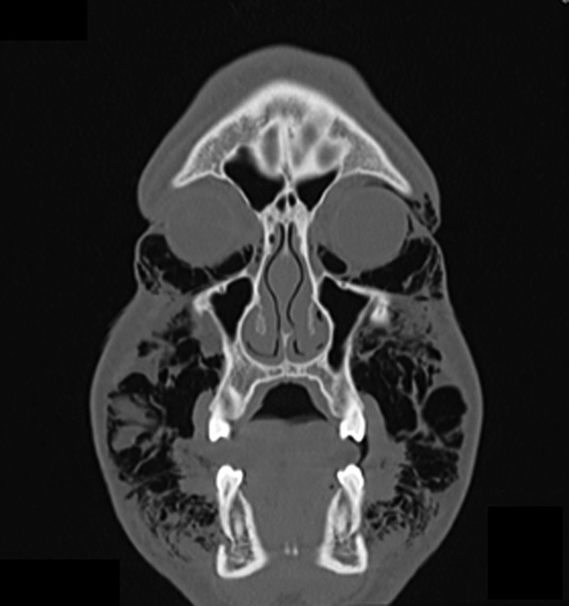
Subcutaneous emphysema is a rare complication in oral surgery. In most cases, it resolves spontaneously. However, air might disperse into deeper facial spaces causing life-threatening complications such as compression of the tracheobronchial tree or the development of pneumomediastinum. Moreover, microorganisms might spread from the oral cavity into deeper spaces. Hence, rapid diagnosis of subcutaneous emphysema is important. Characteristic signs are both a shiftable swelling and crepitation. In this case report, a 30-year-old man, suffering from the Gilles de la Tourette Syndrome, with a distinct subcutaneous emphysema after bilateral surgical wisdom tooth extraction is presented. Induced by a specific motor tic, air accumulated from the periorbital through to the parapharyngeal region. Applying a 10-cm-long Redon drainage tube as air valve, 10 days after wisdom teeth extraction, the patient was asymptomatic with complete resolution of the emphysema.
Published by Oxford University Press and JSCR Publishing Ltd. All rights reserved. © The Author 2015.
Figures


References
-
- Wakoh M, Saitou C, Kitagawa H, Suga K, Ushioda T, Kuroyanagi K. Computed tomography of emphysema following tooth extraction. Dentomaxillofac Radiol 2000;29:201. - PubMed
-
- McKenzie WS, Rosenberg M. Iatrogenic subcutaneous emphysema of dental and surgical origin: a literature review. J Oral Maxillofac Surg 2009;67:1265. - PubMed
-
- Heyman SN, Babayof I. Emphysematous complications in dentistry, 1960–1993: an illustrative case and review of the literature. Quintessence Int 1995;26:535. - PubMed
-
- Suppa A, Marsili L, Di Stasio F, Berardelli I, Roselli V, Pasquini M et al. . Cortical and brainstem plasticity in Tourette syndrome and obsessive-compulsive disorder. Mov Disord 2014;29:1523. - PubMed
-
- Ganos C, Kuhn S, Kahl U, Schunke O, Feldheim J, Gerloff C et al. . Action inhibition in Tourette syndrome. Mov Disord 2014;29:1532. - PubMed
Publication types
LinkOut - more resources
Full Text Sources
Other Literature Sources
