

Targeting the LOX/hypoxia axis reverses many of the features that make pancreatic cancer deadly: inhibition of LOX abrogates metastasis and enhances drug efficacy
- PMID: 26077591
- PMCID: PMC4551344
- DOI: 10.15252/emmm.201404827
Targeting the LOX/hypoxia axis reverses many of the features that make pancreatic cancer deadly: inhibition of LOX abrogates metastasis and enhances drug efficacy
Abstract
Pancreatic ductal adenocarcinoma (PDAC) is one of the leading causes of cancer-related mortality. Despite significant advances made in the treatment of other cancers, current chemotherapies offer little survival benefit in this disease. Pancreaticoduodenectomy offers patients the possibility of a cure, but most will die of recurrent or metastatic disease. Hence, preventing metastatic disease in these patients would be of significant benefit. Using principal component analysis (PCA), we identified a LOX/hypoxia signature associated with poor patient survival in resectable patients. We found that LOX expression is upregulated in metastatic tumors from Pdx1-Cre Kras(G12D/+) Trp53(R172H/+) (KPC) mice and that inhibition of LOX in these mice suppressed metastasis. Mechanistically, LOX inhibition suppressed both migration and invasion of KPC cells. LOX inhibition also synergized with gemcitabine to kill tumors and significantly prolonged tumor-free survival in KPC mice with early-stage tumors. This was associated with stromal alterations, including increased vasculature and decreased fibrillar collagen, and increased infiltration of macrophages and neutrophils into tumors. Therefore, LOX inhibition is able to reverse many of the features that make PDAC inherently refractory to conventional therapies and targeting LOX could improve outcome in surgically resectable disease.
Keywords: animal models of cancer; collagen cross‐linking; lysyl oxidase; pancreatic cancer.
© 2015 Cancer Research UK Beatson Institute. Published under the terms of the CC BY 4.0 license.
Figures
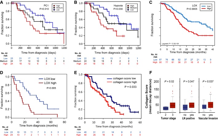
Kaplan–Meier analysis showing cases from the Australian cohort (n = 73) delineated on the basis of expression of a set of hazardous genes, termed PC-1. Patients with the highest expression had a significantly poorer prognosis (red line, median survival: 11.6 months) compared with those patients with the lowest expression (blue line, median survival: 34.4 months, P = 0.01). The black line shows those with medium expression.
Kaplan–Meier analysis showing that cases in the Australian cohort that fell in the highest quartile of a hypoxia signature (red line, median survival: 11.4 months) have significantly decreased survival compared with those in the lowest quartile (blue line, median survival: 25.2 months; P = 0.03).
Kaplan–Meier analysis showing that cases in the APGI cohort (n = 266) with high LOX expression above the 3rd quantile (red line) have significantly decreased survival compared to those with low expression below the 1st quantile (blue line).
Kaplan–Meier analysis showing that cases in the Glasgow cohort with high LOX expression (red line, n = 24) have significantly decreased survival compared with those with low expression (blue line, n = 23; P = 0.005).
Kaplan–Meier analysis showing that fibrillar collagen is significantly associated with reduced survival in human PDAC (20 months vs. 28.3 months, P = 0.033).
Mean decay distance of the second harmonic generation (SHG) signal emitted by human PDAC-associated collagen. Mean decay distance is represented by boxplots showing the second and third quartile of the data with the whiskers indicating the maximum and minimum data points. Outliers are indicated by individual markers. Stage T3 tumors have a higher collagen mean decay distance score compared with Stage T2 tumors (P = 0.02). Lymph node-positive tumors have a higher collagen mean decay distance score vs. lymph node-negative tumors (P = 0.047). Tumors showing vascular invasion have a higher collagen mean decay distance score than non-vascular invasive tumors (P = 0.037).
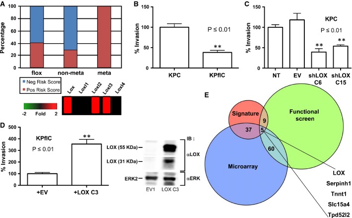
PC-1 signature is predictive of metastatic disease in mouse models of pancreatic cancer. Log-transformed expressions of signature transcripts from mouse tumor microarrays were mean-centered across samples and scaled to unit variance. These values were then multiplied by the matching loading values from the PC-1 signature and summarized for each sample across all transcripts to yield the risk score for that sample.
Inverted invasion assays were performed with PDAC tumor cell lines from KPC and KPflC mice. Tumor cell lines bearing mutant p53R172H (KPC) invade significantly further than tumor cells with deletion of 1 copy of p53 (KPflC) (P ≤ 0.01). Data are shown as the average of four wells + SEM.
Introduction of shRNA targeting Lox into KPC tumor cells significantly inhibits invasion (P ≤ 0.01 by unpaired Student’s t-test). Data are shown as the average of four wells + SEM.
Introduction of exogenous LOX into KPflC tumor cells significantly promotes invasion (left panel, P ≤ 0.01 by unpaired Student’s t-test). LOX expression was assessed by immunoblotting (right panel). Columns indicate the mean of four well and error bars indicate SEM.
Integration of heterogeneous data sets identifies LOX as a therapeutic target in pancreatic cancer. Components of the hazardous PC-1 signature are overlaid with genes found to be overexpressed in a microarray and hits that cause a reduction in viable cell number in an RNAi functional screen. Overlap is shown as a proportional Venn diagram.
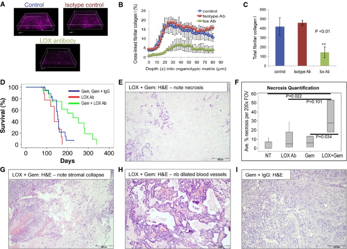
A Representative fluorescence images showing the SHG signal from cross-linked collagen in untreated, LOX antibody-treated or isotype control-treated organotypic cultures of primary fibroblasts and rat tail fibrillar collagen I.
B Chart showing the percentage of cross-linked collagen by depth, based on SHG signal, in organotypic matrices treated as indicated. Error bars indicate SEM.
C Barchart showing total cross-linked collagen in organotypic matrices treated as indicated. The experiment was performed in triplicate with three different zones imaged for 80 μm, at 2.5-μm sections each. Data are shown as total pixels ± SEM.
D Kaplan–Meier (non-parametric) survival curve showing significantly extended tumor-free survival of KPC mice treated with LOX antibody (1 mg/kg i.p., twice weekly) + gemcitabine (100 mg/kg i.p., twice weekly) (green line, n = 17), compared with gemcitabine ± isotype control antibody-treated mice (blue line, n = 22, P = 0.014) or LOX antibody alone (red line, n = 9). Treatment was initiated when mice were 70 days old (randomization was not used when recruiting the mice) and the mice were treated twice weekly. Censored mice did not develop PDAC.
E H&E-stained section of PDAC harvested from LOX + gemcitabine-treated KPC mouse. Note necrosis.
F Boxplot showing quantification of necrosis in H&E sections of PDACs harvested from these mice, as indicated. At least 30 fields of view from at least five mice per cohort were scored.
G–H H&E-stained sections of PDAC harvested from LOX + gemcitabine-treated KPC mice. Note stromal collapse (G) and marked dilation of blood vessels (H).
I Representative H&E image showing a section of PDAC harvested from a control-treated KPC mouse.
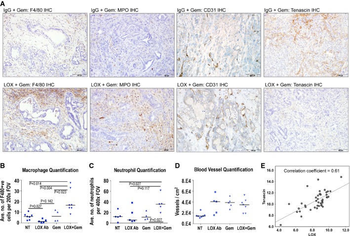
A Outer left panels: Immunohistochemical analysis of macrophage infiltration (by F4/80 staining) in tumors from isotype control + gemcitabine-treated KPC mice vs. LOX antibody + gemcitabine-treated KPC mice. Inner left panels: Immunohistochemical analysis of neutrophil infiltration (by MPO staining) in tumors from isotype control + gemcitabine-treated KPC mice vs. LOX antibody + gemcitabine-treated KPC mice. Inner right panels: Immunohistochemical analysis of tumor vasculature (by CD31 staining) in tumors from isotype control + gemcitabine-treated KPC mice vs. LOX antibody + gemcitabine-treated KPC mice. Outer right panels: Immunohistochemical analysis of tenascin C expression in tumors from isotype control + gemcitabine-treated KPC mice vs. LOX antibody + gemcitabine-treated KPC mice.
B–D Plots showing quantification of macrophage infiltration (B), neutrophil infiltration (C) and tumor vasculature (D) in tumors from KPC mice treated as indicated. At least 30 fields of view from at least four mice per cohort were scored, and scoring was conducted blind. P-values were calculated using Mann–Whitney U-test and median is indicated by horizontal lines.
E Correlation of LOX protein with tenascin C expression in 47 cases of PDAC (Spearman’s rho correlation coefficient = 0.61; P < 0.0001).
References
-
- Adorno M, Cordenonsi M, Montagner M, Dupont S, Wong C, Hann B, Solari A, Bobisse S, Rondina MB, Guzzardo V, et al. A Mutant-p53/Smad complex opposes p63 to empower TGFbeta-induced metastasis. Cell. 2009;137:87–98. - PubMed
-
- Baker AM, Cox TR, Bird D, Lang G, Murray GI, Sun XF, Southall SM, Wilson JR, Erler JT. The role of lysyl oxidase in SRC-dependent proliferation and metastasis of colorectal cancer. J Natl Cancer Inst. 2011;103:407–424. - PubMed
Publication types
MeSH terms
Substances
Grants and funding
LinkOut - more resources
Full Text Sources
Other Literature Sources
Medical
Molecular Biology Databases
Research Materials
Miscellaneous
