. 2015 Jun 16:12:119.
doi: 10.1186/s12974-015-0335-3.

Interleukin-22 is increased in multiple sclerosis patients and targets astrocytes

Affiliations

Interleukin-22 is increased in multiple sclerosis patients and targets astrocytes

Guillaume Perriard et al. J Neuroinflammation. .

Abstract

Background: Increasing evidences link T helper 17 (Th17) cells with multiple sclerosis (MS). In this context, interleukin-22 (IL-22), a Th17-linked cytokine, has been implicated in blood brain barrier breakdown and lymphocyte infiltration. Furthermore, polymorphism between MS patients and controls has been recently described in the gene coding for IL-22 binding protein (IL-22BP). Here, we aimed to better characterize IL-22 in the context of MS.

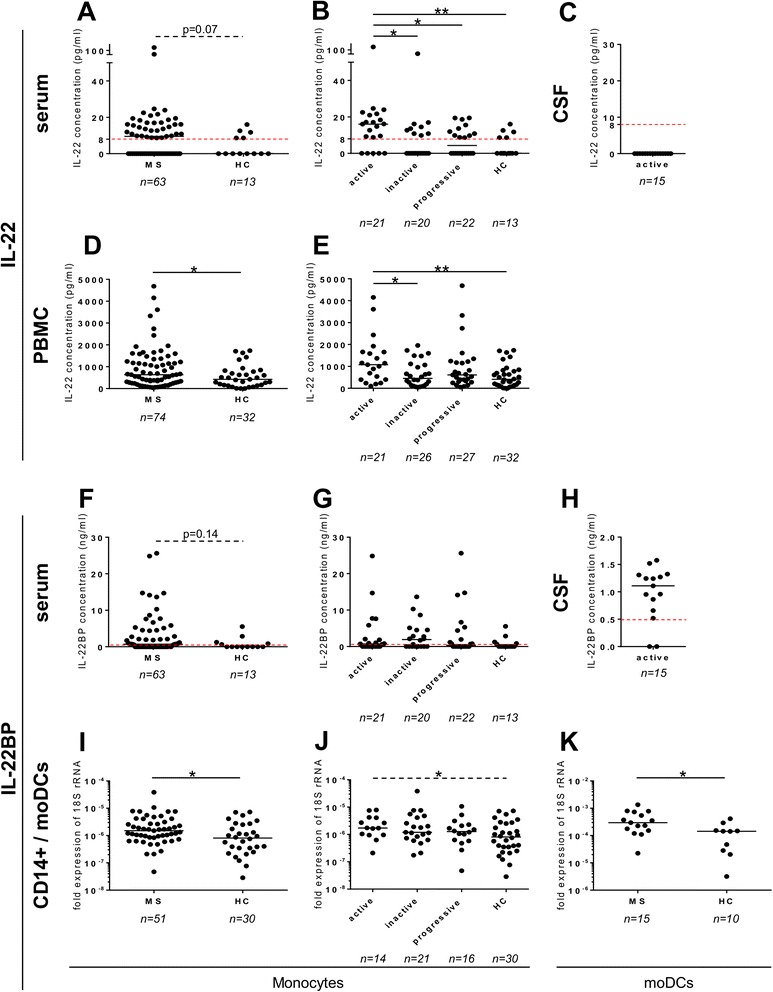
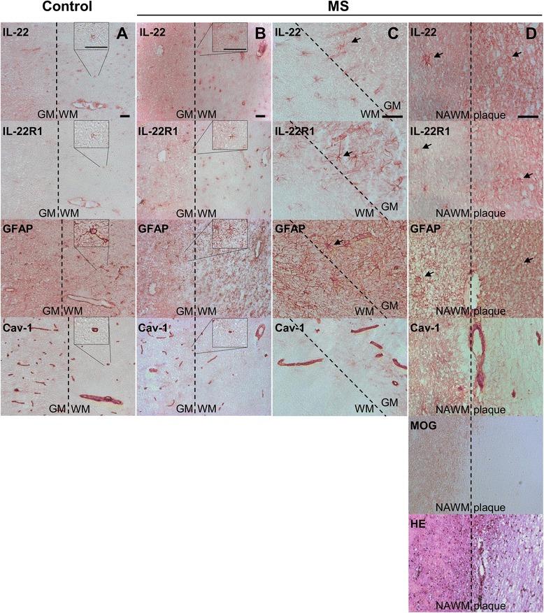
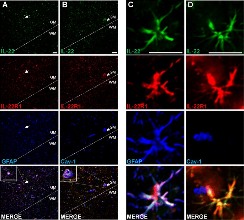
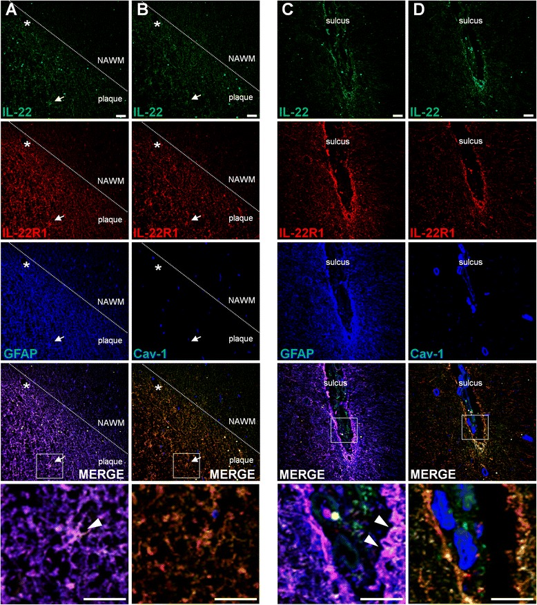
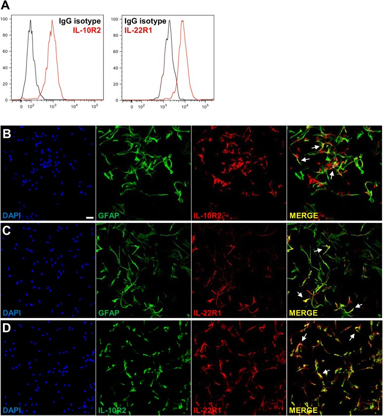
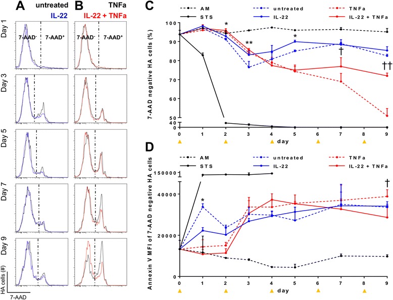
Methods: IL-22 and IL-22BP expressions were assessed by ELISA and qPCR in the following compartments of MS patients and control subjects: (1) the serum, (2) the cerebrospinal fluid, and (3) immune cells of peripheral blood. Identification of the IL-22 receptor subunit, IL-22R1, was performed by immunohistochemistry and immunofluorescence in human brain tissues and human primary astrocytes. The role of IL-22 on human primary astrocytes was evaluated using 7-AAD and annexin V, markers of cell viability and apoptosis, respectively.

Results: In a cohort of 141 MS patients and healthy control (HC) subjects, we found that serum levels of IL-22 were significantly higher in relapsing MS patients than in HC but also remitting and progressive MS patients. Monocytes and monocyte-derived dendritic cells contained an enhanced expression of mRNA coding for IL-22BP as compared to HC. Using immunohistochemistry and confocal microscopy, we found that IL-22 and its receptor were detected on astrocytes of brain tissues from both control subjects and MS patients, although in the latter, the expression was higher around blood vessels and in MS plaques. Cytometry-based functional assays revealed that addition of IL-22 improved the survival of human primary astrocytes. Furthermore, tumor necrosis factor α-treated astrocytes had a better long-term survival capacity upon IL-22 co-treatment. This protective effect of IL-22 seemed to be conferred, at least partially, by a decreased apoptosis.

Conclusions: We show that (1) there is a dysregulation in the expression of IL-22 and its antagonist, IL-22BP, in MS patients, (2) IL-22 targets specifically astrocytes in the human brain, and (3) this cytokine confers an increased survival of the latter cells.


Figures

Fig. 1
Fig. 1
IL-22BP and IL-22 are increased in MS patients as compared to healthy controls. The IL-22 and IL-22BP expressions were assessed by ELISA (a–h) and qPCR (i–k) in the serum (a, b and f, g), CSF (c, h), isolated monocytes (i, j), and moDCs (k) and supernatant of SEB-stimulated PBMC for 18 h (d, e) isolated from MS patients and healthy controls. Each dot represents a patient. The bars represent the median. Dashed red lines represent the detection limit. Active: clinically active MS patients, inactive: clinically inactive MS patients, progressive: primary and secondary progressive MS patients. Differences among the four groups were significant with Kruskal-Wallis test (b, e). Unpaired non-parametric Mann–Whitney test was used to compare groups two-by-two. *P < 0.05, **P < 0.01
Fig. 2
Fig. 2
IL-22 and IL-22R1 are expressed in human brain. IL-22, IL-22R1, GFAP, and Caveolin-1 immunohistochemistry peroxidase stainings of brain tissue sections of control (a) and MS (bd) patients. MOG and HE stainings were used to detect MS demyelinating plaques (d). All four pictures belonging to one column (a, b, c, or d) were always immediately adjacent to each other. Pictures a, b, and c were taken at areas at the border between GM and NAWM, whereas pictures in d were taken from the same location at the edge between NAWM and a plaque. Inserts in columns a and b represent a threefold magnification of the selected area. Arrow: astrocyte-like pattern. a study patient B-C2, b and d study patient B-MS3, c study patient B-MS5 (Table 2). Scale bar, 50 μm (a, b: ×20, c, d ×40). GM: gray matter, NAWM: normal appearing white matter, WM: white matter. Representative pictures obtained from the observations of seven control and five MS autopsy samples
Fig. 3
Fig. 3
Specific expression of IL-22 and its receptor, IL-22R1, by astrocytes in the brains of control patients without MS and suffering from other neurological diseases. Laser scanning confocal microscopy observations were performed in brain tissue autopsies (Basel cohort). Brain autopsy labeled for IL-22 (first panel, green), IL-22R1 (second panel, red), and Caveolin-1 or GFAP (third panel, blue). Merged images are depicted as composite images in the lower panel (colocalization of IL-22R1 and GFAP appears in yellow and triple colocalization of IL-22, IL-22R1, and GFAP in white). Inserts are a ×10 zoom of the selected area (a, b, lowest panels). Images a and b were taken on autopsied brain tissue from control patient B-C6 and c and d from control patient B-C1 (Table 2). Arrows in a point at astrocytes, and stars in b at blood vessels. Bars, 50 μm. GM: gray matter, WM: white matter. Representative pictures obtained from the observations of seven control autopsy samples
Fig. 4
Fig. 4
Strong expression of IL-22 and its receptor, IL-22R1, in the plaques of MS brains. Immunofluorescence of human brain tissue sections stained for IL-22 (green), IL-22R1 (red), and Cav-1 or GFAP (blue) in MS brain tissue autopsies. Images were taken from patient B-MS3 patient (Table 2) in a plaque located in the subcortical WM (a, b) and in a plaque located in the subpial GM (c, d). The lowest panels depict a ×5 magnification of the white squares displayed in the above panels. Arrow: astrocytes, star: blood vessels. Arrowheads point at triple IL-22/IL-22R1/GFAP colocalization (a, c). Bars, 50 μm. NAWM: normal appearing white matter. Representative pictures obtained from the observations of five MS autopsy samples
Fig. 5
Fig. 5
Colocalization of both subunits of IL-22 receptor on primary astrocytes. a Flow cytometry experiments show the expression of both subunits of the IL-22 receptor, IL-10R2, and IL-22R1 (in red), on human primary astrocytes (HA) as compared to their isotype control counterparts (in black). bd By immunofluorescent confocal microscopy, there is a colocalization of GFAP (green) with IL-10R2 (red) (b) and with IL-22R1 (red) (c) as well as of IL-10R2 (green) with IL-22R1 (red) (d). The colocalization appears is depicted in yellow (arrows, MERGE). DAPI is represented in blue and not included in MERGE overlay. Bars, 50 μm
Fig. 6
Fig. 6
Enhanced survival of IL-22-treated primary astrocytes in insulting conditions. Primary astrocyte cells (HA) were cultured in 24-well plates starting at day −1 and were treated every second day starting at day 0 with astrocyte medium (AM)—representing the optimal culture medium for astrocytes—or with RPMI (poor medium, hereafter referred to as untreated), with or without addition of the following: IL-22 alone, TNFα alone, IL-22 and TNFα together, or still staurosporine (STS) alone, the latter representing a potent inducer of apoptosis. Cells were stained with 7-AAD and Annexin V and analyzed by flow cytometry to assess their survival and apoptotic status. Histograms represent 7-AAD profile of untreated versus IL-22 (a) and of TNFα versus IL-22 + TNFα (b). Survival profile analysis was performed by flow cytometry by following the frequency of living cells (i.e., 7-AAD-negative cells, Fig. 6 c) and, among those that were not dead (7-AAD-negative cells), by following the proportion of those surviving cells but which underwent apoptosis (d). Each dot represents the median of six replicates, except for AM and STS conditions (three replicates). Orange arrows indicate treatment renewal. Considering STS treatment, 7-AAD kinetic was stopped after 4 days as all recovered cells were dead at this time point.*comparison of IL-22-treated versus untreated cells; †comparison of TNFα- versus TNFα + IL-22-treated cells. The vertical bars determine the 75th percentile of the median. Significance was calculated with unpaired non-parametric Mann–Whitney test. * or †P < 0.05, ** or ††P < 0.01

References

    1. Compston A, Coles A. Multiple sclerosis. Lancet. 2008;372:1502–17. doi: 10.1016/S0140-6736(08)61620-7. - DOI - PubMed
    1. Sospedra M, Martin R. Immunology of multiple sclerosis. Annu Rev Immunol. 2005;23:683–747. doi: 10.1146/annurev.immunol.23.021704.115707. - DOI - PubMed
    1. Maddur MS, Miossec P, Kaveri SV, Bayry J. Th17 cells: biology, pathogenesis of autoimmune and inflammatory diseases, and therapeutic strategies. Am J Pathol. 2012;181:8–18. doi: 10.1016/j.ajpath.2012.03.044. - DOI - PubMed
    1. Luchtman DW, Ellwardt E, Larochelle C, Zipp F. IL-17 and related cytokines involved in the pathology and immunotherapy of multiple sclerosis: current and future developments. Cytokine Growth Factor Rev. 2014;25:403–13. doi: 10.1016/j.cytogfr.2014.07.013. - DOI - PubMed
    1. Sie C, Korn T, Mitsdoerffer M. Th17 cells in central nervous system autoimmunity. Exp Neurol. 2014;262:18–27. doi: 10.1016/j.expneurol.2014.03.009. - DOI - PubMed

Publication types

MeSH terms

LinkOut - more resources