. 2015 Jun 16:5:11385.
doi: 10.1038/srep11385.

A G-quadruplex-binding compound showing anti-tumour activity in an in vivo model for pancreatic cancer

Affiliations

A G-quadruplex-binding compound showing anti-tumour activity in an in vivo model for pancreatic cancer

Stephan A Ohnmacht et al. Sci Rep. .

Abstract

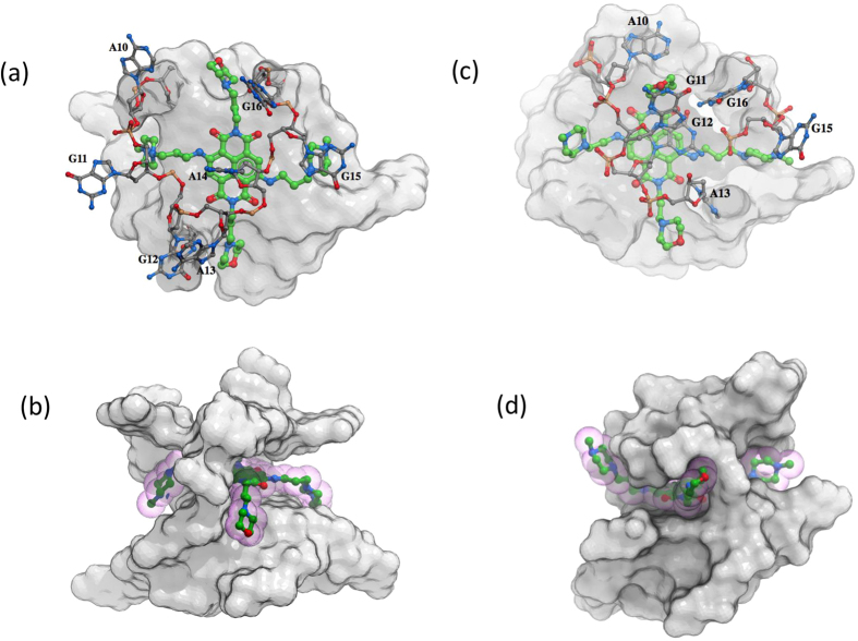
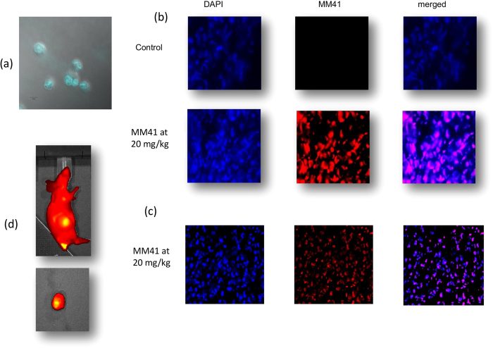
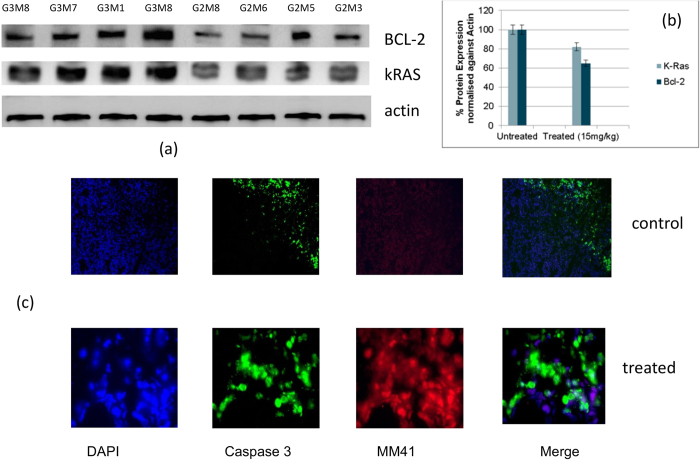
We report here that a tetra-substituted naphthalene-diimide derivative (MM41) has significant in vivo anti-tumour activity against the MIA PaCa-2 pancreatic cancer xenograft model. IV administration with a twice-weekly 15 mg/kg dose produces ca 80% tumour growth decrease in a group of tumour-bearing animals. Two animals survived tumour-free after 279 days. High levels of MM41 are rapidly transported into cell nuclei and were found to accumulate in the tumour. MM41 is a quadruplex-interactive compound which binds strongly to the quadruplexes encoded in the promoter sequences of the BCL-2 and k-RAS genes, both of which are dis-regulated in many human pancreatic cancers. Levels of BCL-2 were reduced by ca 40% in tumours from MM41-treated animals relative to controls, consistent with BCL-2 being a target for MM41. Molecular modelling suggests that MM41 binds to a BCL-2 quadruplex in a manner resembling that previously observed in co-crystal structures with human telomeric quadruplexes. This supports the concept that MM41 (and by implication other quadruplex-targeting small molecules) can bind to quadruplex-forming promoter regions in a number of genes and down-regulate their transcription. We suggest that quadruplexes within those master genes that are up-regulated drivers for particular cancers, may be selective targets for compounds such as MM41.


Figures

Figure 1
Figure 1. The structure of MM41.
Figure 2
Figure 2. Visualisations of the binding of a MM41 molecule to a BCL-2 promoter quadruplex, taken from the molecular modelling study.
(a) Top view of the quadruplex with the MM41 molecule involved in π-π interactions with the terminal G-quartet. (b) Side view of the docked complex. The four side-chain arms of MM41 penetrate into the four grooves of the quadruplex; however, the side-chains are not long enough to be involved in direct interactions with the backbone. (c) Top view of the final snapshot from the MD simulations. Base G12 forms π-stacking interactions with the chromophore of MM41, which is then sandwiched between G12 and the top G-quartet. (d) Side view of the final snapshot from the MD simulations illustrating the rearrangement of loop-2 nucleotides, which now interact with the side chains of MM41. The drug is shown as green sticks while loop-2 surface is shown in cpk colours.
Figure 3
Figure 3
(a) Efficacy of MM41 in subcutaneous tumor xenografts, evaluated in terms of tumour growth over time ± SEM. Control mice are compared to groups dosed with 10 and 15 mg/kg respectively, administered twice daily. The change in tumour volume for the group dosed with 15 mg/kg of MM41 is statistically significant (Student’s t-test) relative to the control group with P = 0.026 (n = 6). (b) Average mouse weight over time for the three experimental groups of mice. (c) Kaplan-Meier survival plot for the three groups of mice. Note that cessation of particular experiments at day 40 (see text) accounts for the zero survival of 2/3 groups at this time-point.
Figure 4
Figure 4
(a) Confocal image of MIA PaCa-2 cells following a 30 min exposure to MM41. The fluorescence of the compound is seen concentrated in the cell nuclei. (b) Images (x 40) of tumour tissue from control and MM41-treated MIA PaCa-2 xenografts, showing DAPI-stained and MM41 fluorescent images, together with merged images showing superposition of DAPI and MM41 staining. The images are of sections close to the tumour surface. (c) Images (x 20) showing DAPI and MM41 staining, together with the merged image, for sections taken from the centre of a treated tumour. (d) (LHS) IVIS whole-body fluorescence image, taken at the MM41 emission wavelength, showing that MM41 is concentrated in the tumour, which is on the flank of the animal. A substantial amount of MM41 is shown to be retained at the site of administration. An image of the extracted tumour is also shown.
Figure 5
Figure 5
(a) Western blots for BCL-2 and k-RAS proteins, and actin as a control. The four LHS columns represent blots from four untreated mice, and the four RHS ones are from four MM41-treated (at 15 mg/kg). All mice were taken at the termination of the therapeutic experiments. (b) Plot of the averaged changes in BCL-2 and k-RAS proteins, normalised against the actin blots. (c) Images (X 10) of caspase immuno-staining in sections from control and MM41-treated tumours.

References

    1. Vincent A., Herman J., Schulick R., Hruban R. H. & Goggins M. Pancreatic cancer. Lancet 378, 607–620 (2011). - PMC - PubMed
    1. Matthaei H., Semaan. A. & Hruban R. H. The genetic classification of pancreatic neoplasia. J. Gastroenterol. (2015) 10.1007/s00535-015-1037-4. - DOI - PubMed
    1. http://www.cancer.org/cancer/pancreaticcancer/detailedguide/pancreatic-c.... Date of access 06/02/2015.
    1. http://www.cancerresearchuk.org/cancer-info/cancerstats/types/pancreas/i... of access 06/02/2015.
    1. Sant M. et al. EUROCARE-4. Survival of cancer patients diagnosed in 1995–1999. Results and commentary. Eur. J. Cancer 45, 931–991 (2009). - PubMed

Publication types

MeSH terms