. 2015 Jun 15;308(12):L1237-44.
doi: 10.1152/ajplung.00042.2015. Epub 2015 Apr 10.

Remodeling of alveolar septa after murine pneumonectomy

Affiliations

Remodeling of alveolar septa after murine pneumonectomy

Alexandra B Ysasi et al. Am J Physiol Lung Cell Mol Physiol. .

Abstract

In most mammals, removing one lung (pneumonectomy) results in the compensatory growth of the remaining lung. In mice, stereological observations have demonstrated an increase in the number of mature alveoli; however, anatomic evidence of the early phases of alveolar growth has remained elusive. To identify changes in the lung microstructure associated with neoalveolarization, we used tissue histology, electron microscopy, and synchrotron imaging to examine the configuration of the alveolar duct after murine pneumonectomy. Systematic histological examination of the cardiac lobe demonstrated no change in the relative frequency of dihedral angle components (Ends, Bends, and Junctions) (P > 0.05), but a significant decrease in the length of a subset of septal ends ("E"). Septal retraction, observed in 20-30% of the alveolar ducts, was maximal on day 3 after pneumonectomy (P < 0.01) and returned to baseline levels within 3 wk. Consistent with septal retraction, the postpneumonectomy alveolar duct diameter ratio (Dout:Din) was significantly lower 3 days after pneumonectomy compared to all controls except for the detergent-treated lung (P < 0.001). To identify clumped capillaries predicted by septal retraction, vascular casting, analyzed by both scanning electron microscopy and synchrotron imaging, demonstrated matted capillaries that were most prominent 3 days after pneumonectomy. Numerical simulations suggested that septal retraction could reflect increased surface tension within the alveolar duct, resulting in a new equilibrium at a higher total energy and lower surface area. The spatial and temporal association of these microstructural changes with postpneumonectomy lung growth suggests that these changes represent an early phase of alveolar duct remodeling.

Keywords: electron microscopy; microstructure; regeneration; surface tension.


Figures

Fig. 1.
Fig. 1.
Morphometry of the postpneumonectomy cardiac lobe. A: serial histological sections of the cardiac lobe were obtained on various days after pneumonectomy (day 0), stained with hematoxylin and eosin, and imaged for morphometry. The planar sections were thresholded (inset) and the tissue density calculated as a percent of planar area. B: Similar histological sections of the cardiac lobe were examined for E, B, and J frequency. Defined as “ends,” “bends,” and “junctions” (inset, highlighted definitions of EBJ) (6), respectively, each feature was expressed as a percent of the total features counted. Differences over the 22-day time course were not significant (P > 0.05). Error bars reflect 1 SD. N = 4–6 mice per time point.
Fig. 2.
Fig. 2.
Histology of the cardiac lobe in control (A and C) and postpneumonectomy day 3 mice (B and D). The cellular lung in control (A) and postpneumonectomy day 3 (B) cardiac lobe were stained with Sirius red. Normal septa were thin and projected into the alveolar duct (A, ellipses). Postpneumonectomy day 3 sections frequently demonstrated “plump” septa that appeared to be retracted away from the duct lumen (B, arrows). C and D: lungs of control and postpneumonectomy day 3 mice were decellularized and the cardiac lobe stained with Verhoeff-van Gieson stain. The dilated alveolar ducts demonstrated areas of condensed matrix (D, arrows), suggesting both cellular and matrix alterations after pneumonectomy. Bar = 100 μm.
Fig. 3.
Fig. 3.
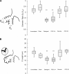
Morphometry of the alveolar duct in the murine cardiac lobe. Serial sections of the cardiac lobe were obtained in various conditions. A: width of the alveolar duct was expressed as a ratio between outer diameter (distance between outer walls, Dout) and inner diameter of the duct (distance between septal tips, Din). B: E length was measured as the greatest length along the axis of the septa. These parameters were compared in cardiac lobes from nonsurgical controls, sham thoracotomy mice, detergent-treated mice, postpneumonectomy day 3 (POD 3) mice, areas of the cardiac lobe remote from known growth areas (remote) and postpneumonectomy day 22 (POD 22) mice. Error bars = 1 SD. POD 3 duct diameters were indistinguishable from detergent-treated mice (P > 0.05), but significantly different from the other controls (**P < 0.001). N = 3–5 mice per condition.
Fig. 4.
Fig. 4.
Morphometry of the cardiac lobe alveolar ducts between pneumonectomy (day 0) and postpneumonectomy day 22. Serial sections of the cardiac lobe were obtained in various conditions. A: when all of the alveolar ducts were evaluated, variability was noted on day 3 after pneumonectomy (*P < 0.05). B: when the duct measurements at all time points were combined and the most dilated 20% plotted as a function of time after pneumonectomy, most of the dilated ducts were identified on day 3 after pneumonectomy. C: similarly, septal length (E) decreased on day 3 after pneumonectomy (*P < 0.05). The difference in septal length E was also significant between day 3 and day 12 after pneumonectomy (**P < 0.01). D: in contrast, other structural features, such as B angle, were unchanged (P > 0.05). N = 3–6 mice per time point.
Fig. 5.
Fig. 5.
Corrosion casting and scanning electron microscopy of the control (A and B) and postpneumonectomy lung (C and D). A and B: in control lungs, corrosion casts of the cardiac lobe demonstrated the typical appearance of capillaries within the normal alveoli. Bar = 200 μm. C and D: in contrast, the cardiac lobe of lungs casted 3 days after pneumonectomy demonstrate areas of high density or “clumped” capillaries (arrows). Bar = 200 μm.
Fig. 6.
Fig. 6.
Synchrotron images of vascular casts obtained from the cardiac lobe of control (A and C) and postpneumonectomy (B and D) mice. Sequential images (N = 10) at 650-nm intervals were digitally recombined for presentation purposes. A and C: in control mice, there were narrow septa (ellipses) with only rare evidence of capillary clumps. B and D: in contrast, postpneumonectomy day 35 mice demonstrated many more areas with dense capillaries (arrows) consistent with septal retraction (representative image is shown). C: high-resolution imaging of the control lung demonstrated alveolar walls generally appearing to be one vessel thick. D: in contrast, walls of the alveolar duct frequently demonstrated dense vascular areas consistent with septal retraction (arrows).
Fig. 7.
Fig. 7.
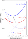
Numerical simulation of the effect of increased surface tension in the alveolar duct. Total lung energy E is the sum of tissue energy U and surface energy ∫ γdS. In the normal lung (red line), surface area S takes the value for which total energy is minimum (red dot). After pneumonectomy (blue line), an increase in surface energy, assumed to be equivalent to the detergent-treated lung (2), results in a shift of the curve (gray arrow). In a hypothetical lung with retracted septa, the minimum total energy (blue dot) occurs at a smaller surface area and larger total energy. Adapting the quadratic energy difference model of Wilson (39), we assumed a fixed lung volume (80% total lung capacity). A, air; D, detergent.

References

    1. Ackermann M, Houdek JP, Gibney BC, Ysasi AB, Wagner W, Belle JM, Schittny JC, Enzmann F, Tsuda A, Mentzer SJ, Konerding MA. Sprouting and intussusceptive angiogenesis in postpneumonectomy lung growth: mechanisms of alveolar neovascularization. Angiogenesis 17: 541–551, 2013. - PMC - PubMed
    1. Bachofen H, Gehr P, Weibel ER. Alterations of mechanical-properties and morphology in excised rabbit lungs rinsed with a detergent. J Appl Physiol 47: 1002–1010, 1979. - PubMed
    1. Befera NT, Badea CT, Johnson GA. Comparison of 4D-MicroSPECT and MicroCT for murine cardiac function. Mol Imaging Biol 16: 235–245, 2014. - PMC - PubMed
    1. Bennett RA, Ysasi AB, Belle JM, Wagner W, Konerding MA, Blainey P, Pyne S, Mentzer SJ. Laser microdissection of the alveolar duct for single-cell genomic analysis. Front Oncol 4: 260, 2014. - PMC - PubMed
    1. Butler J, Loring SH, Patz S, Tsuda A, Yablonskiy DA, Mentzer SJ. Evidence for adult lung growth in humans. N Engl J Med 367: 244–247, 2012. - PMC - PubMed

Publication types

LinkOut - more resources