. 2015 Jun;7(1):49-54.
doi: 10.1007/s12593-014-0167-x. Epub 2015 Jan 8.

Neural Anatomy of the Anterolateral Thigh Flap

Affiliations

Neural Anatomy of the Anterolateral Thigh Flap

Suriya Luenam et al. J Hand Microsurg. 2015 Jun.

Abstract

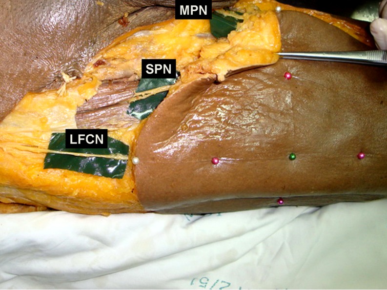
The anterolateral thigh (ALT) flap is one of the commonly used sensate flaps for intra-oral, hand, and foot reconstruction. The objective of this study was to describe the anatomic location of the sensory nerves supplying the ALT flap in relation to the surface landmarks and with the vascular pedicles. The dissections were carried out in 28 embalmed specimens. An axial line from the anterior superior iliac spine to the superolateral border of the patella and two circles with radii of 5 and 10 cm centered on the midpoint of the former line were used for the surface landmarks. At the intersection point of the axial line and the 10-cm circle, the main lateral femoral cutaneous nerve (LFCN) and its anterior branch were located within 1 and 2.4 cm, respectively. At the intersection point of the axial line and the 5-cm circle, the anterior branch of the LFCN was located within 2.8 cm. The anterior branch of the LFCN can be detected within 3 cm from the central perforator pedicle in all specimens. The posterior branch of the LFCN, superior perforator nerve, and median perforator nerve were found in more variable locations. The findings from our study provide additional information for clinical use in the planning of sensate ALT flap harvest.

Keywords: Anterolateral thigh flap; Lateral femoral cutaneous nerve; Neural anatomy; Sensate flap.


Figures

Fig. 1
Fig. 1
Surface landmarks were established, and ALT flaps with a skin territory of 10 and 20 cm in diameter were designed
Fig. 2
Fig. 2
Anatomical dissection of a left anterolateral thigh flap. LFCN lateral femoral cutaneous nerve, SPN superior perforator nerve, MPN median perforator nerve
Fig. 3
Fig. 3
The distance correlation of the nerves with the surface landmarks and vascular pedicles was measured
Fig. 4
Fig. 4
The locations of the LFCN, SPN, and MPN
Fig. 5
Fig. 5
The number of dermal innervations of the LFCN was higher in the lower half of the flap
Fig. 6
Fig. 6
Illustration of the surface landmarks and nerve locations. CL common LFCN, AL anterior branch of LFCN, P vascular pedicle

References

    1. Kimata Y, Uchiyama K, Ebihara S, et al. Comparison of innervated and noninnervated free flaps in oral reconstruction. Plast Reconstr Surg. 1999;104:1307–1313. doi: 10.1097/00006534-199910000-00010. - DOI - PubMed
    1. Hong JP, Kim EK. Sole reconstruction using anterolateral thigh perforator free flaps. Plast Reconstr Surg. 2007;119:186–193. doi: 10.1097/01.prs.0000244856.98170.9c. - DOI - PubMed
    1. Ducic I, Hung V, Dellon AL. Innervated free flaps for foot reconstruction: a review. J Reconstr Microsurg. 2006;22:433–442. doi: 10.1055/s-2006-947698. - DOI - PubMed
    1. Shridharani SM, Magarakis M, Stapleton SM. Breast sensation after breast reconstruction: a systematic review. J Reconstr Microsurg. 2010;26:303–310. doi: 10.1055/s-0030-1249313. - DOI - PubMed
    1. Netscher D, Armenta AH, Meade RA, et al. Sensory recovery of innervated and non-innervated radial forearm free flaps: functional implications. J Reconstr Microsurg. 2000;16:179–185. doi: 10.1055/s-2000-7549. - DOI - PubMed

LinkOut - more resources