

Syringocystadenocarcinoma Papilliferum in the Perianal Area
- PMID: 26078737
- PMCID: PMC4464026
- DOI: 10.1159/000381940
Syringocystadenocarcinoma Papilliferum in the Perianal Area
Abstract
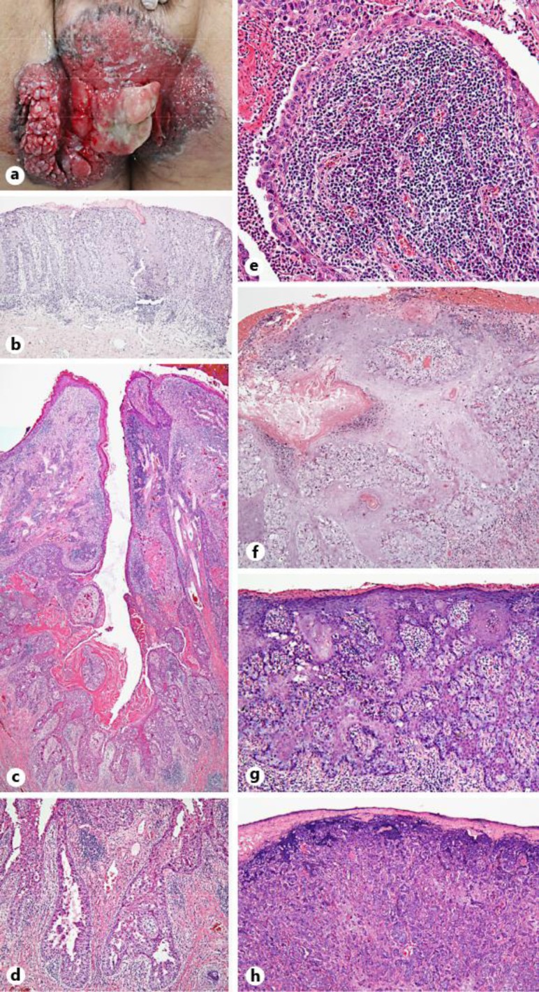
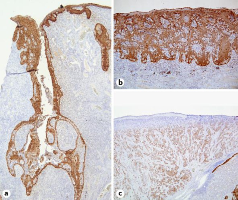
Syringocystadenocarcinoma papilliferum (SCACP) is a very rare cutaneous adnexal neoplasm. SCACP presents histologic variability, and it is difficult to establish the diagnosis from a punch biopsy. SCACP has an overall configuration similar to that of syringocystadenoma papilliferum (SCAP). When we diagnose SCACP, the histologic features of SCAP can be contributing and immunohistochemical staining is useful. Our case shows the histologic variability of SCACP and the pitfalls of a punch biopsy for the diagnosis of SCACP.
Keywords: Adenocarcinoma; Cutaneous adnexal neoplasm; Lymph node metastasis; Pagetoid spread; Perianal area; Perianal syringocystadenocarcinoma papilliferum; Syringocystadenocarcinoma papilliferum.
Figures
References
-
- Castillo L, Moreno A, Tardio JC. Syringocystadenocarcinoma papilliferum in situ: report of a case with late recurrence. Am J Dermatopathol. 2014;36:348–352. - PubMed
-
- Kazakov DV, Requena L, Kutzner H, Fernandez-Figueras MT, Kacerovska D, Mentzel T, Schwabbauer P, Michal M. Morphologic diversity of syringocystadenocarcinoma papilliferum based on a clinicopathologic study of 6 cases and review of the literature. Am J Dermatopathol. 2010;32:340–347. - PubMed
-
- Zhang YH, Wang WL, Rapini RP, Torres-Cabala C, Prieto VG, Curry JL. Syringocystadenocarcinoma papilliferum with transition to areas of squamous differentiation: a case report and review of the literature. Am J Dermatopathol. 2012;34:428–433. - PubMed
-
- Liegl B, Leibl S, Gogg-Kamerer M, Tessaro B, Horn LC, Moinfar F. Mammary and extramammary Paget's disease: an immunohistochemical study of 83 cases. Histopathology. 2007;50:439–447. - PubMed
-
- Langner C, Ott A. Syringocystadenocarcinoma papilliferum in situ originating from the perianal skin. APMIS. 2009;117:148–150. - PubMed
Publication types
LinkOut - more resources
Full Text Sources
Other Literature Sources
