. 2015 May 23;6(7):623-31.
doi: 10.7150/jca.11291. eCollection 2015.

Platycodin-D Induced Autophagy in Non-Small Cell Lung Cancer Cells via PI3K/Akt/mTOR and MAPK Signaling Pathways

Affiliations

Platycodin-D Induced Autophagy in Non-Small Cell Lung Cancer Cells via PI3K/Akt/mTOR and MAPK Signaling Pathways

Ruolin Zhao et al. J Cancer. .

Abstract

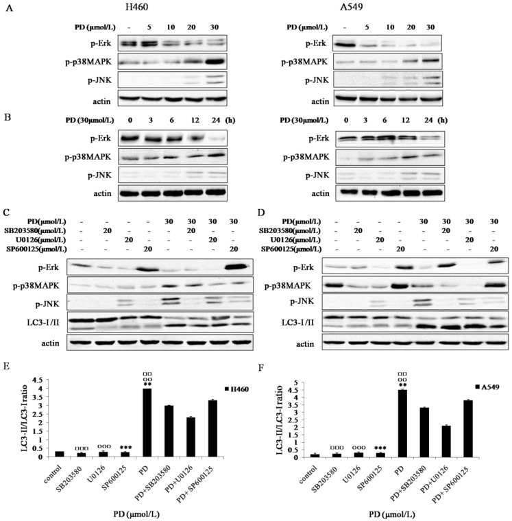
Platycodin-D (PD) is an effective triterpene saponin extracted from the root of Platycodon grandiflorum which has been used clinically to treat pulmonary diseases in traditional Chinese medicine. Recently, it has been reported that PD has anti-tumor effects in various cancer models through the induction of apoptosis. However, whether PD induces autophagy in both cell lines and its molecular mechanisms have not been elucidated. Here, our present study confirmed that PD induced autophagy in both NCI-H460 and A549 cells via up-regulating the expression levels of Atg-3, Atg-7 and Beclin-1. Meanwhile, PD contributed to the up-regulation of LC3-II at both protein and mRNA levels. Further detection of the PI3K/Akt/mTOR signaling pathway compared to LY294002 (PI3K kinase inhibitor), RAP (mTOR kinase inhibitor) and insulin (an activator of PI3K/Akt/mTOR signaling pathway) showed that PD induced autophagy through inhibiting the pathway at p-Akt (Ser473), p-p70S6K (Thr389) and p-4EBP1 (Thr37/46) in both cell lines. Moreover, the examination of MAPK signaling pathway showed that PD treatment increased the phosphorylation of JNK and p38 MAPK, while decreased the phosphorylation of Erk1/2 in both cell lines. Additionally, the effects assessed with a panel of pharmacologic inhibitors, including U0126 (Erk1/2 kinase inhibitor), SP600125 (JNK kinase inhibitor) and SB203580 (p38 MAPK kinase inhibitor) suggested that the activation of JNK and p38 MAPK participated in PD-induced autophagy. Taken together, these findings suggested that PD induced autophagy in NCI-H460 and A549 cells through inhibiting PI3K/Akt/mTOR signaling pathway and activating JNK and p38 MAPK signaling pathways. Therefore, PD may be an alternative compound for NSCLC therapy.

Keywords: MAPK signaling pathways; PI3K/Akt/mTOR; Platycodin-D; autophagy; non-small cell lung cancer.


Conflict of interest statement

Conflict of Interest: The authors have declared that no conflict of interest exists.

Figures

Figure 1
Figure 1
PD induced morphological changes of NCI-H460 and A549 cells. (A) NCI-H460 and A549 cells treated with PD at various concentrations of 0, 10, 20, and 30 μmol/L, respectively. After 24 h treatment, cells stained with Gimesa were observed using phase-contrast microscopy (×400). (B) NCI-H460 and A549 cells were exposed to 0, 20 and 30 μmol/L of PD for 24h followed by observation using a transmission electron microscope (TEM). Numerous autophagosomes with typical double-layer membranes containing organelle remnants were highlighted by arrows.
Figure 2
Figure 2
Effect of PD on inducing autophagy in NCI-H460 and A549 cells. (A and B) NCI-H460 and A549 cells treated with 0, 5, 10, 20 and 30 µmol/L of PD for 24 h or 30 µmol/L of PD for 0, 3, 6, 12 and 24 h were analyzed by western-blot with antibodies against LC3-I/II, Beclin-1, Atg-3 and Atg-7. (C and D) Densitometry analysis of LC3-II level relative to actin was performed. (E) The mRNA expression level of LC3-II induced by PD in both cell lines was detected by Quantitative reverse transcription-PCR analysis. Representative results of three independent experiments are shown. β-actin was used as a loading control. Error bars, SD; *, P<0.05; **, P<0.01, ***, P<0.001 versus control values.
Figure 3
Figure 3
Effect of PD on PI3K/Akt/mTOR/p70s6k/4EBP1 signaling pathway in NCI-H460 and A549 cells. (A and B) NCI-H460 and A549 cells treated with 0, 5, 10, 20 and 30 µmol/L of PD for 24 h or 30 µmol/L of PD for 0, 3, 6, 12 and 24 h were analyzed by western-blot with antibodies against p-Akt (Ser473), p-p70S6K(Thr389), p-4EBP1 (Thr37/46), Akt, p70S6K and 4EBP1. (C and D) NCI-H460 and A549 cells treated with 30 µmol/L of PD, 10 µmol/L of LY294002 or 10 µmol/L of Rapmycin for 24 h were analyzed by western-blot with antibodies against p-Akt (Ser473), p-p70S6K (Thr389), p-4EBP1 (Thr37/46) and LC3-І /II. (E and F) Cells treated with 30 µmol/L of PD for 24 h followed by treatment with or without 200 nmol/L of insulin for 30 min were analyzed by western-blot for the expression levels of p-Akt (Ser473), p-p70S6K (Thr389) and LC3-І/II. (G and H) Densitometry analysis of LC3-II level relative to actin was performed in both two cell lines.○, □: P<0.05; and ○○, □□: P<0.01 versus PD + RAP values; *, △: P<0.05; and **, △△: P<0.01 versus PD + LY294002 values. △△△: P<0.001 Insulin versus PD + Insulin values; **: P<0.01 PD versus control values. Representative results of three independent experiments are shown. β-actin was used as a loading control. Error bars, SD.
Figure 4
Figure 4
Effect of PD on MAPK signaling pathways in NCI-H460 and A549 cells. (A and B) NCI-H460 and A549 cells treated with 0, 5, 10, 20 and 30 µmol/L of PD for 24 h or 30 µmol/L of PD for 0, 3, 6, 12 and 24 h were analyzed by western blot with antibodies against p-Erk1/2, p-JNK and p-p38 MAPK. (C and D) NCI-H460 and A549 cells treated with 20 µmol/L of SB203580, 20 µmol/L of U0126 or 20 µmol/L of SP600125 for 4 h followed by treatment with or without 30 µmol/L of PD for 24 h were analyzed by western blot with antibodies against p-Erk 1/2 , p-JNK, p-p38 MAPK and LC3-І /II. (E and F) Densitometry analysis of LC3-II levels relative to LC3-І in NCI-H460 and A549 cells was performed. Representative results of three independent experiments are shown. β-actin was used as a loading control. Error bars, SD; □□: P<0.01 and □□□: P<0.001: versus PD + SB203580 values; ○○: P<0.01 and ○○○: P<0.001 versus PD + U0126 values; **: P<0.01 and ***: P<0.001 versus PD + SP600125 values.
Figure 5
Figure 5
A schematic model of the molecular mechanisms associated with PD-induced autophagy in NCI-H460 and A549 cells. According to this model, when NCI-H460 and A549 cells were exposed to PD, the phagosome converted to double-layered membranes of autophagosomes through increasing expression levels of Atg proteins including Beclin-1, Atg-3 and Atg-7 as well as leading to the conversion of LC3-I to LC3-II. The inhibition of PI3K/Akt/mTOR signaling pathway and activation of JNK and p38 MAPK signaling pathways contributed to the accumulation of LC3-II, which were the possible upstream signaling pathways of PD-induced autophagy. Once the autophagosome was developed, its maturation was complete upon fusion with lysosome to form autophagolysosome. Eventually, programmed cell death was induced by PD. The schematic model compiled with the results and conclusions of this study (→: Stimulatory Modification, ⊥: Inhibitory Modification).

References

    1. Chun J, Joo EJ, Kang M. et al. Platycodin D induces anoikis and caspase-mediated apoptosis via p38 MAPK in AGS human gastric cancer cells. Journal of cellular biochemistry. 2013;114(2):456–470. - PubMed
    1. Yu JS, Kim AK. Platycodin D induces apoptosis in MCF-7 human breast cancer cells. Journal of medicinal food. 2010;13(2):298–305. - PubMed
    1. Kim MO, Moon DO, Choi YH. et al. Platycodin D induces mitotic arrest in vitro, leading to endoreduplication, inhibition of proliferation and apoptosis in leukemia cells. International journal of cancer Journal international du cancer. 2008;122(12):2674–2681. - PubMed
    1. Janku F, McConkey DJ, Hong DS. et al. Autophagy as a target for anticancer therapy. Nature reviews Clinical oncology. 2011;8(9):528–539. - PubMed
    1. Chen S, Rehman SK, Zhang W. et al. Autophagy is a therapeutic target in anticancer drug resistance. Biochimica et biophysica acta. 2010;1806(2):220–229. - PubMed