

Cytomegalovirus Uveitis with Hypopyon Mimicking Bacterial Endophthalmitis
- PMID: 26078897
- PMCID: PMC4442280
- DOI: 10.1155/2015/489813
Cytomegalovirus Uveitis with Hypopyon Mimicking Bacterial Endophthalmitis
Abstract
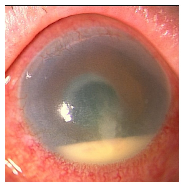
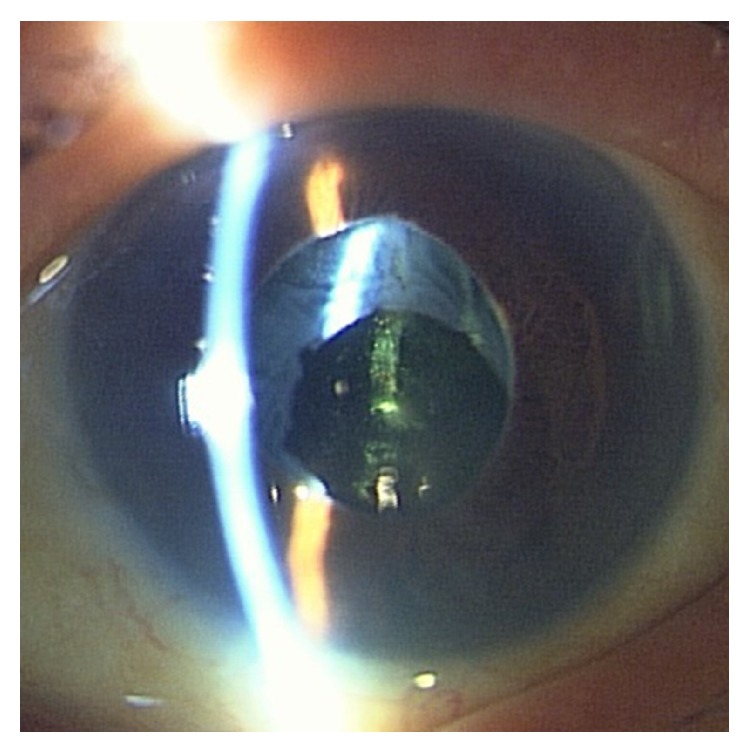
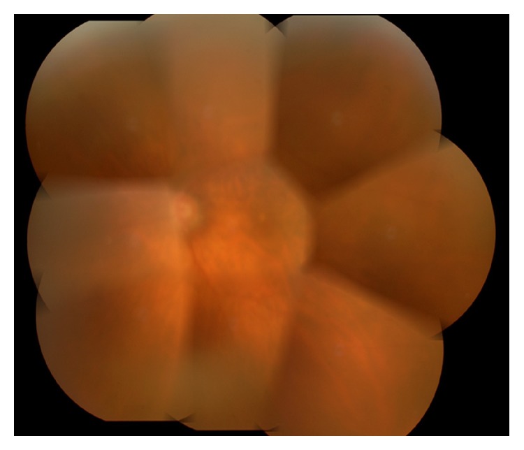
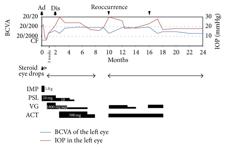
We report an 83-year-old immune-competent female with unilateral endophthalmitis extraordinarily caused by cytomegalovirus (CMV). Since she was suspected of suffering possible bacterial endophthalmitis, she was referred to our hospital. At the first visit, hypopyon in the anterior chamber and the opacity of vitreous body were observed in the left eye. The best-corrected visual acuity (BCVA) of the left eye was counting fingers and the intraocular pressure (IOP) was 20 mmHg. Bacterial and fungus culture of the aqueous humor revealed no infection. However, the density of corneal endothelial cell was less than the measurable range and CMV was detected by PCR of the aqueous humor. She was immune-competent and the data indicated neither systemic infections nor diseases. Systemic valganciclovir and corticosteroid were administered. After that, hypopyon in the anterior chamber and the opacity of vitreous body of the left eye were improved, and the BCVA of the left eye was 20/200 one year after the first visit. However, the inflammation of the anterior chamber recurred accompanied by elevated IOP after the discontinuance of administering valganciclovir. CMV-induced uveitis accompanied with hypopyon is quite rare. Therefore, it can be easily misdiagnosed as bacterial endophthalmitis.
Figures
Similar articles
-
Cytomegalovirus Retinitis with Hypopyon Similar to Bacterial Endophthalmitis in a Patient with non-Hodgkin's Lymphoma.Ocul Immunol Inflamm. 2022 Oct-Nov;30(7-8):2017-2018. doi: 10.1080/09273948.2021.1944647. Epub 2021 Jul 2. Ocul Immunol Inflamm. 2022. PMID: 34213982
-
Acute endophthalmitis and hyphema mimicking pink hypopyon associated with ocular toxocariasis: A case report.Am J Ophthalmol Case Rep. 2021 Aug 10;23:101188. doi: 10.1016/j.ajoc.2021.101188. eCollection 2021 Sep. Am J Ophthalmol Case Rep. 2021. PMID: 34430757 Free PMC article.
-
Cytomegalovirus as a cause of anterior uveitis in immunocompetent patients.Ophthalmology. 2007 Jul;114(7):1358-62. doi: 10.1016/j.ophtha.2006.09.035. Epub 2007 Feb 12. Ophthalmology. 2007. PMID: 17296229
-
Endogenous Serratia marcescens endophthalmitis with dark hypopyon: case report and review.Surv Ophthalmol. 2001 Nov-Dec;46(3):259-68. doi: 10.1016/s0039-6257(01)00263-6. Surv Ophthalmol. 2001. PMID: 11738433 Review.
-
Listeria monocytogenes endophthalmitis - case report and review of risk factors and treatment outcomes.BMC Infect Dis. 2016 Jul 16;16:332. doi: 10.1186/s12879-016-1680-2. BMC Infect Dis. 2016. PMID: 27424034 Free PMC article. Review.
Cited by
-
Combined endophthalmitis and orbital cellulitis in patients with corona virus disease (COVID-19).J Ophthalmic Inflamm Infect. 2021 Sep 15;11(1):27. doi: 10.1186/s12348-021-00258-y. J Ophthalmic Inflamm Infect. 2021. PMID: 34523045 Free PMC article.
-
Mimickers of anterior uveitis, scleritis and misdiagnoses- tips and tricks for the cornea specialist.J Ophthalmic Inflamm Infect. 2024 Apr 10;14(1):14. doi: 10.1186/s12348-024-00396-z. J Ophthalmic Inflamm Infect. 2024. PMID: 38594487 Free PMC article. Review.
References
LinkOut - more resources
Full Text Sources
Other Literature Sources
