. 2015:2015:543818.
doi: 10.1155/2015/543818. Epub 2015 May 20.

Altered Expression of NF- κ B and SP1 after Exposure to Advanced Glycation End-Products and Effects of Neurotrophic Factors in AGEs Exposed Rat Retinas

Affiliations

Altered Expression of NF- κ B and SP1 after Exposure to Advanced Glycation End-Products and Effects of Neurotrophic Factors in AGEs Exposed Rat Retinas

Guzel Bikbova et al. J Diabetes Res. 2015.

Abstract

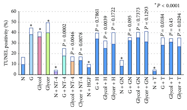
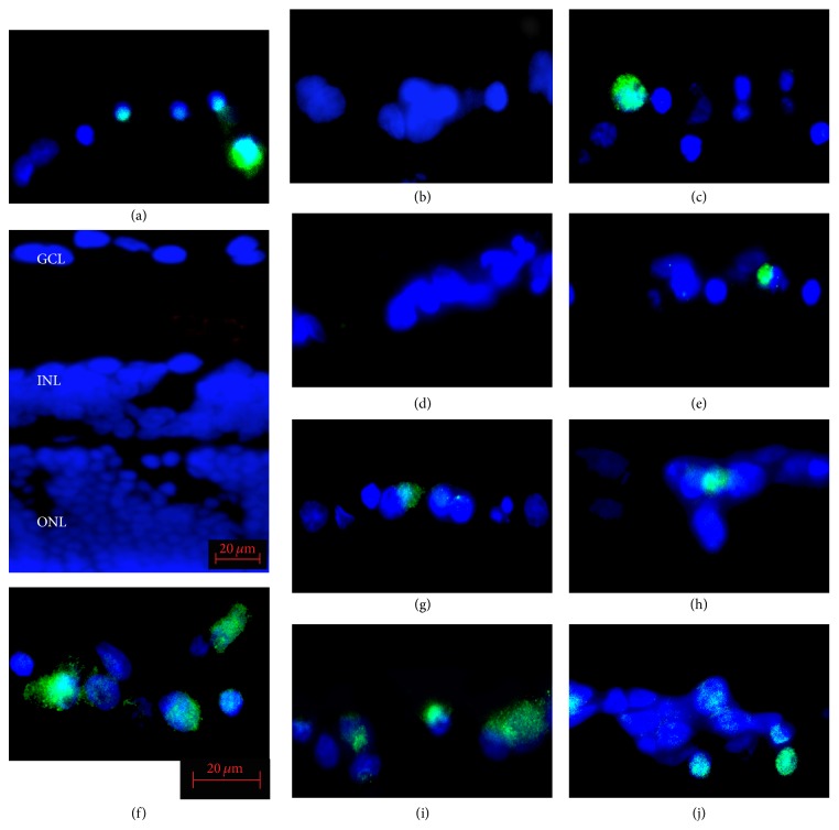
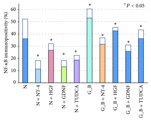
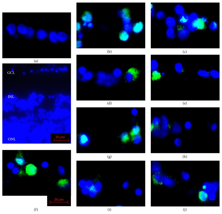
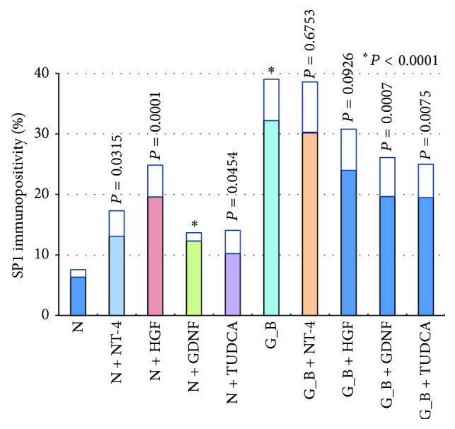
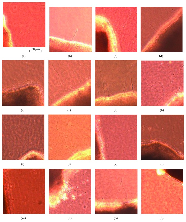
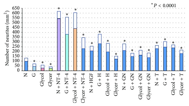
To determine the effect of advanced glycation end-products (AGEs) on neurite regeneration, and also to determine the regenerative effects of different neurotrophic factors (NTFs) on rat retinal explants, the retinas of SD rats were cultured in three-dimensional collagen gels and incubated in 6 types of media: (1) serum-free control culture media; (2) 100 μg/mL AGEs-BSA media; (3) AGEs-BSA + 100 ng/mL neurotrophin-4 (NT-4) media; (4) AGEs-BSA + 100 ng/mL hepatocyte growth factor media; (5) AGEs-BSA + 100 ng/mL glial cell line-derived neurotrophic factor media; or (6) AGEs-BSA + 100 µM tauroursodeoxycholic acid media. After 7 days, the number of regenerating neurites was counted. The explants were immunostained for nuclear factor-κB (NF-κB) and specificity protein 1 (SP1). Statistical analyses were performed by one-way ANOVA. In retinas incubated with AGEs, the numbers of neurites were fewer than in control. All of the NTFs increased the number of neurites, and the increase was more significant in the NT-4 group. The number of NF-κB and SP1 immunopositive cells was higher in retinas exposed to AGEs than in control. All of the NTFs decreased the number of NF-κB immunopositive cells but did not significantly affect SP1 expression. These results demonstrate the potential of the NTFs as axoprotectants in AGEs exposed retinal neurons.


Figures

Figure 1
Figure 1
Graph showing the ratio of TUNEL-positive cells to all cells in the GCL of retinal explants. N, serum-free media; G, glucose-AGE-BSA; Glycol, glycolaldehyde-AGE-BSA; Glycer, glyceraldehyde-AGE-BSA; NT-4, neurotrohin-4; H, hepatocyte growth factor; GN, glial cell line-derived neurotrophic factor; T, tauroursodeoxycholic acid.
Figure 2
Figure 2
Representative photomicrographs of NF-κB immunopositive cells in the ganglion cell layer (GCL) of isolated rat retinas. In retinas cultured in glucose-AGE-BSA (f), the number of immunopositive cells in the GCL is significantly higher than in serum-free control media (a). In AGEs exposed retinas supplemented with NT-4 (glucose-AGE-BSA + NT-4) (g), with HGF (glucose-AGE-BSA + HGF) (h), with GDNF (glucose-AGE-BSA + GDNF) (i), and with TUDCA (glucose-AGE-BSA + TUDCA) (j) the number of NF-κB immunopositive cells is fewer than that in AGEs exposed retinas without neurotrophic factors. The blue staining shows the DAPI-stained nuclei. Panels (b), (c), (d), and (e) are pictures of the serum-free media supplemented with NT-4 (b), HGF (c), GDNF (d), and TUDCA (e). INL, inner nuclear layer; ONL, outer nuclear layer. Bar = 20 μm.
Figure 3
Figure 3
Graph showing the ratio of NF-κB immunopositive cells to all cells in the GCL of retinal explants. N, serum-free media; G_B, glucose-AGE-BSA; Glycol, glycolaldehyde-AGE-BSA; Glycer, glyceraldehyde-AGE-BSA; NT-4, neurotrohin-4; HGF, hepatocyte growth factor; GDNF, glial cell line-derived neurotrophic factor; TUDCA, tauroursodeoxycholic acid.
Figure 4
Figure 4
Representative photomicrographs of SP1 immunopositive cells in the ganglion cell layer (GCL) of isolated rat retinas. In retinas cultured in glucose-AGE-BSA (f), the number of immunopositive cells in the GCL is significantly higher than in serum-free control media (a). In AGEs exposed retinas supplemented with NT-4 (glucose-AGE-BSA + NT-4) (g), with HGF (glucose-AGE-BSA + HGF) (h), with GDNF (glucose-AGE-BSA + GDNF) (i), and with TUDCA (glucose-AGE-BSA + TUDCA) (j) the number of SP1 immunopositive cells is higher than that in AGEs exposed retinas without neurotrophic factors. The blue signals show the DAPI-stained nuclei. Panels (b), (c), (d), and (e) show the representative pictures of the serum-free media incubated with NT-4 (b), HGF (c), GDNF (d), and TUDCA (e). INL, inner nuclear layer; ONL, outer nuclear layer. Bar = 20 μm.
Figure 5
Figure 5
Graph showing the ratio of SP1 immunopositive cells to all cells in the GCL of retinal explants. N, serum free media, G_B, glucose-AGE-BSA; Glycol, glycolaldehyde-AGE-BSA; Glycer, glyceraldehyde-AGE-BSA; NT-4, neurotrohin-4; HGF, hepatocyte growth factor, GDNF, glial cell line-derived neurotrophic factor; TUDCA, tauroursodeoxycholic acid.
Figure 6
Figure 6
Representative photographs of regenerating neurites. Regenerating neurites are seen under phase-contrast microscopy. In the control serum-free media (a) neurites with normal length are present. In retinas cultured in glucose-AGE-BSA (b), glyceraldehyde-AGE-BSA (c), and glycolaldehyde-AGE-BSA (d), the neurites were shorter, and the numbers of neurites were fewer. In AGEs exposed retinas supplemented with NT-4 (glucose-AGE-BSA + NT-4 (e), glyceraldehyde-AGE-BSA + NT-4 (f), and glycolaldehyde-AGE-BSA + NT-4 (g)), the neurites are longer and thicker, and the number of neurites are higher even than in serum-free control media (a). In AGEs exposed retinas supplemented with HGF, GDNF, and TUDCA (glucose-AGE-BSA + HGF (h), glyceraldehyde-AGE-BSA + HGF (i), and glycolaldehyde-AGE-BSA + HGF (j)), (glucose-AGE-BSA + GDNF (k), glyceraldehyde-AGE-BSA + GDNF (l), and glycolaldehyde-AGE-BSA + GDNF (m)), (glucose-AGE-BSA + TUDCA (n), glyceraldehyde-AGE-BSA + TUDCA (o), and glycolaldehyde-AGE-BSA + TUDCA (p)), the neurites are longer and thicker than in AGEs exposed retinas.
Figure 7
Figure 7
Graph showing the number of regenerating neurites in all groups. N, serum-free media, G, glucose-AGE-BSA; Glycol, glycolaldehyde-AGE-BSA; Glycer, glyceraldehyde-AGE-BSA; NT-4, neurotrohin-4; H, hepatocyte growth factor; GN, glial cell line-derived neurotrophic factor; T, tauroursodeoxycholic acid.

References

    1. Ruderman N. B., Williamson J. R., Brownlee M. Glucose and diabetic vascular disease. The FASEB Journal. 1992;6(11):2905–2914. - PubMed
    1. Wautier J.-L., Wautier M.-P., Schmidt A.-M., et al. Advanced glycation end products (AGEs) on the surface of diabetic erythrocytes bind to the vessel wall via a specific receptor inducing oxidant stress in the vasculature: a link between surface associated AGEs and diabetic complications. Proceedings of the National Academy of Sciences of the United States of America. 1994;91(16):7742–7746. doi: 10.1073/pnas.91.16.7742. - DOI - PMC - PubMed
    1. Bikbova G., Oshitari T., Yamamoto S. Neurite regeneration in adult rat retinas exposed to advanced glycation end-products and regenerative effects of neurotrophin-4. Brain Research. 2013;1534:33–45. doi: 10.1016/j.brainres.2013.08.027. - DOI - PubMed
    1. Schmidt A. M., Hori O., Brett J., Yan S. D., Wautier J.-L., Stern D. Cellular receptors for advanced glycation end products: implications for induction of oxidant stress and cellular dysfunction in the pathogenesis of vascular lesions. Arteriosclerosis, Thrombosis, and Vascular Biology. 1994;14(10):1521–1528. doi: 10.1161/01.atv.14.10.1521. - DOI - PubMed
    1. Vlassara H., Li Y. M., Imani F., et al. Identification of galectin-3 as a high-affinity binding protein for advanced glycation end products (AGE): a new member of the AGE-receptor complex. Molecular Medicine. 1995;1(6):634–646. - PMC - PubMed

Publication types

MeSH terms

LinkOut - more resources