Observational Study
. 2015:2015:729217.
doi: 10.1155/2015/729217. Epub 2015 May 20.

Achalasia--An Autoimmune Inflammatory Disease: A Cross-Sectional Study

Affiliations
Observational Study

Achalasia--An Autoimmune Inflammatory Disease: A Cross-Sectional Study

J Furuzawa-Carballeda et al. J Immunol Res. 2015.

Abstract

Idiopathic achalasia is a disease of unknown etiology. The loss of myenteric plexus associated with inflammatory infiltrates and autoantibodies support the hypothesis of an autoimmune mechanism. Thirty-two patients diagnosed by high-resolution manometry with achalasia were included. Twenty-six specimens from lower esophageal sphincter muscle were compared with 5 esophagectomy biopsies (control). Immunohistochemical (biopsies) and flow cytometry (peripheral blood) analyses were performed. Circulating anti-myenteric autoantibodies were evaluated by indirect immunofluorescence. Herpes simplex virus-1 (HSV-1) infection was determined by in situ hybridization, RT-PCR, and immunohistochemistry. Histopathological analysis showed capillaritis (51%), plexitis (23%), nerve hypertrophy (16%), venulitis (7%), and fibrosis (3%). Achalasia tissue exhibited an increase in the expression of proteins involved in extracellular matrix turnover, apoptosis, proinflammatory and profibrogenic cytokines, and Tregs and Bregs versus controls (P < 0.001). Circulating Th22/Th17/Th2/Th1 percentage showed a significant increase versus healthy donors (P < 0.01). Type III achalasia patients exhibited the highest inflammatory response versus types I and II. Prevalence of both anti-myenteric antibodies and HSV-1 infection in achalasia patients was 100% versus 0% in controls. Our results suggest that achalasia is a disease with an important local and systemic inflammatory autoimmune component, associated with the presence of specific anti-myenteric autoantibodies, as well as HSV-1 infection.


Figures

Figure 1
Figure 1
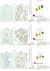
Proteins involved in extracellular matrix turnover in achalasia. (a) Representative photomicrograph from a control and an idiopathic achalasia tissue in which an abundant inflammatory infiltrate is observed. Original magnification was ×200. (b) Histological findings in biopsies from achalasia patients. (c) Immunoreactive cells. Original magnification was ×320. (d) Results are expressed as mean (yellow line), median (black line), and 5th/95th percentiles of MMP-9+ cells. (e) Immunohistochemistry for TIMP-1 in control and achalasia sections. Arrows depict immunoreactive cells. Original magnification was ×320. (f) Results are expressed as mean (black line), median (yellow line), and 5th/95th percentiles of TIMP-1+ cells.
Figure 2
Figure 2
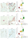
Proinflammatory/antifibrogenic cytokine expression in achalasia. Immunohistochemistry for (a) IL-22, (c) IL-17, and (e) CD4/IFN-γ T cells in control and achalasia sections. Arrows depict immunoreactive cells. Original magnification was ×320. Results are expressed as mean (yellow line), median (black line), and 5th/95th percentiles of (b) IL-22+, (d) IL-17+, and (f) CD4/IFN-γ + T cells.
Figure 3
Figure 3
Anti-inflammatory/profibrogenic cytokine expression in achalasia. Immunohistochemistry for (a) TGF-β1, (c) IL-4, and (e) IL-13 cells in control and achalasia sections. Arrows depict immunoreactive cells. Original magnification was ×320. Results are expressed as mean (yellow line), median (black line), and 5th/95th percentiles of (b) TGF-β1+, (d) IL-4+, and (f) IL-13+ cells.
Figure 4
Figure 4
Regulatory cells and apoptosis in achalasia. Immunohistochemistry for (a) CD25/Foxp3 T cells, (c) CD20/IL-10 B cells, and (e) FAS cells in control and achalasia sections. Arrows depict immunoreactive cells. Original magnification was ×320. Results are expressed as mean (yellow line), median (black line), and 5th/95th percentiles of (b) CD25/Foxp3+ T cells, (d) CD20/IL-10+ B cells, and (f) FAS+ cells.
Figure 5
Figure 5
Circulating CD4+ T-cell subsets in achalasia. Relative percentage of circulating (a) Th22, (b) Th17, (c) Th2, (d) Th1, and (e) regulatory T cells. Results are expressed as mean (yellow line), median (black line), and 5th/95th percentiles of T cells.
Figure 6
Figure 6
Anti-myenteric plexus antibodies and antigenicity. (a) Indirect immunofluorescence of monkey intestinal tissue containing Auerbach's plexus. Arrows show positive fluorescent nuclei and cytoplasm of cells that were recognized by circulating anti-myenteric plexus autoantibodies from achalasia patients (right panel). (b) Profile of neuronal antigens (antigens on membrane strips). Arrow in upper strip shows circulating autoantibodies against PNMA2 (Ma2/Ta) antigen. Arrow in lower strip shows circulating antibodies against recoverin antigen.
Figure 7
Figure 7
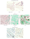
HSV-1 identification. (a) In situ hybridization. Arrows depict achalasia tissue with viral infection. (b) In situ RT-PCR for active replication of HSV-1. Arrows show antigen detection of HSV-1 infected cells in a skin lesion (positive control) and achalasia tissue. Original magnification was ×200. (c) Immunohistochemistry for antigen HSV-1 detection. Arrows depict immunoreactive cells. Original magnification was ×320.
Figure 8
Figure 8
Proposed model of achalasia pathophysiology. (1) Active or latent infectious insult in achalasia is strongly suggested by several studies. Some neurotropic viruses such as the herpes family of viruses have predilection for squamous epithelium and may cause ganglion cell damage that is limited to the esophagus. (2) Some individuals with genetic predisposition will develop an aggressive inflammatory response. (3) At very early stage of the disease, it is possible that inflammatory infiltrates may be predominantly composed of Th1, Th2, and regulatory cell subsets (Tregs, Bregs, and plasmacytoid dendritic cells (pDCs)). (4) Repair of tissue after injury requires orchestrated coordination of several cell types and biosynthetic processes and is coordinated by an interacting group of pro- and anti-inflammatory cytokines, fibrous extracellular matrix (ECM) proteins to replace lost or damaged tissue, and products of metabolism such as oxygen radicals. The most prominent profibrogenic cytokines are TGFβ, IL-4, and IL-13. ECM also mediates cellular crosstalk and does so in two ways. Newly deposited ECM is then rebuilding over time to emulate normal tissue. Matrix proteinases and their inhibitors (TIMPs) also are important, both during wound repair, tissue remodeling, and fibrosis. (2′, 5) If steps 1–4 happen repeatedly, chronic infection, only those individuals with genetic predisposition to develop a long-lasting autoinflammatory response will progress to develop the disease (loss of peripheral tolerance). Thus, autoinflammatory infiltrates are predominantly composed of Th22, Th17, and regulatory subpopulations. (6) Degeneration and significant loss of nerve fibers, associated with autoinflammatory infiltrates of the myenteric plexus, provide evidence of an immune mediated destruction of the inhibitory neurons, not only by necrosis but also by apoptosis (Fas/FasL overexpression). (7) Autoimmune etiology of achalasia is further supported by the presence of anti-myenteric autoantibodies in sera. (8) Pathophysiologically, achalasia is cause by autoinflammation, degeneration of nerves in the esophagus, plexitis, abnormalities in microvasculature, ganglionitis, and finally by the loss of inhibitory ganglion in the myenteric plexus.

References

    1. Boeckxstaens G. E. Novel mechanism for impaired nitrergic relaxation in achalasia. Gut. 2006;55(3):304–305. doi: 10.1136/gut.2005.078402. - DOI - PMC - PubMed
    1. Bruley des Varannes S., Chevalier J., Pimont S., et al. Serum from achalasia patients alters neurochemical coding in the myenteric plexus and nitric oxide mediated motor response in normal human fundus. Gut. 2006;55(3):319–326. doi: 10.1136/gut.2005.070011. - DOI - PMC - PubMed
    1. Mayberry J. F. Epidemiology and demographics of achalasia. Gastrointestinal Endoscopy Clinics of North America. 2001;11(2):235–247. - PubMed
    1. Boeckxstaens G. E., Zaninotto G., Richter J. E. Achalasia. The Lancet. 2014;383(9911):83–93. doi: 10.1016/s0140-6736(13)60651-0. - DOI - PubMed
    1. Pandolfino J. E., Kwiatek M. A., Nealis T., Bulsiewicz W., Post J., Kahrilas P. J. Achalasia: a new clinically relevant classification by high-resolution manometry. Gastroenterology. 2008;135(5):1526–1533. doi: 10.1053/j.gastro.2008.07.022. - DOI - PMC - PubMed

Publication types

MeSH terms

LinkOut - more resources