. 2015 Jul;46(7):948-56.
doi: 10.1016/j.humpath.2015.03.017. Epub 2015 Apr 15.

PAX8 expression in ovarian surface epithelial cells

Affiliations

PAX8 expression in ovarian surface epithelial cells

Emily Adler et al. Hum Pathol. 2015 Jul.

Abstract

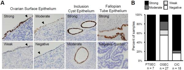
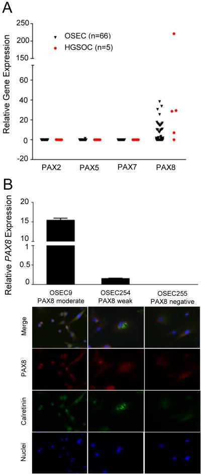
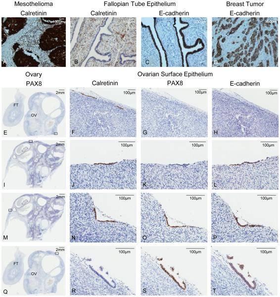
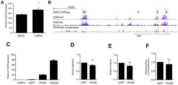
High-grade serous ovarian carcinoma (HGSOC) is usually diagnosed at a late stage and is associated with poor prognosis. Understanding early stage disease biology is essential in developing clinical biomarkers to detect HGSOC earlier. While recent studies indicate that HGSOCs arise from fallopian tube secretory epithelial cells, a considerable body of evidence suggests that HGSOC can also arise from ovarian surface epithelial cells (OSECs). PAX8 is overexpressed in HGSOCs and expressed in fallopian tube secretory epithelial cells, but there are conflicting reports about PAX8 expression in OSECs. The purposes of this study were to comprehensively characterize PAX8 expression in a large series of OSECs and to investigate the role of PAX8 in early HGSOC development. PAX8 protein expression was analyzed in the OSECs of 27 normal ovaries and 7 primary OSEC cultures using immunohistochemistry and immunofluorescent cytochemistry. PAX8 messenger RNA expression was quantified in 66 primary OSEC cultures. Cellular transformation was evaluated in OSECs expressing a PAX8 construct. PAX8 was expressed by 44% to 71% of OSECs. Calretinin and E-cadherin were frequently coexpressed with PAX8. Expression of PAX8 in OSECs decreased cellular migration (P = .028), but had no other effects on cellular transformation. In addition, PAX8 expression was significantly increased (P = .003) in an in vitro stepwise model of neoplastic transformation. In conclusion, PAX8 is frequently expressed by OSECs, and endogenous levels of PAX8 expression are non-transforming. These data indicate that in OSECs, PAX8 expression may represent a normal state and that OSECs may represent an origin of HGSOCs.

Keywords: CMYC; Fallopian tube epithelial cells; High-grade serous ovarian carcinoma; Inclusion cyst; Ovarian surface epithelium; PAX8.


Figures

Figure 1
Figure 1
PAX8 expression in normal ovarian surface epithelium. (A) PAX8 expression was evaluated in 27 normal ovaries using immunohistochemistry. PAX8 expression ranged from strong to negative in OSECs and was strong, moderate or negative in CIC epithelial cells. FTSECs stained positive for PAX8. Examples of normal OSEC morphology on the ovarian surface are indicated with a black arrowhead. Tissue sections are shown at 100x magnification, negative control shown at 40x. (B) Graphical illustration of the range and proportion of tissues with PAX8 expression in primary OSEC, CIC, and FTSEC tissue. OSEC = ovarian surface epithelial cell, CIC = cortical inclusion cyst, FTSEC = fallopian tube secretory epithelial cell.
Figure 2
Figure 2
PAX8 protein and mRNA expression is correlated in OSECs. (A) Boxplot comparing relative PAX gene expression in primary OSEC cultures (black triangles), normalized to B-Actin and GAPDH and calibrated to a single OSEC line (OSEC250). HGSOC cell lines are shown as red circles. (B) OSEC cultures stained for PAX8 displayed PAX8 moderate expression (left panel and bar), weak expression (middle panel and bar), or negative expression (right panel and bar). Expression of PAX8 detected by immunofluorescence correlated with mRNA abundance. PAX8 moderate and weak mRNA expression was significantly different (P < 0.0001, one way ANOVA). 84% of OSEC9 and OSEC254 cells were PAX8 positive, and 48% of OSEC255 cells were weakly positive for PAX8. All cultures were calretinin positive by immunofluorescence using an anti-calretinin antibody. All images were taken at 200x magnification. OSEC = ovarian surface epithelial cell, HGSOC = high-grade serous ovarian carcinoma.
Figure 3
Figure 3
PAX8, calretinin, and E-cadherin expression in normal ovarian surface epithelium. (A-D) Controls are shown in the top panel. (A) mesothelioma with positive calretinin staining, (B) fallopian tube epithelium negative for calretinin and positive for E-cadherin expression (negative E-cadherin staining seen in stroma), (C) breast tumor specimen positive for E-cadherin expression. (E, I, M, Q) A section of normal ovary stained for PAX8. The black rectangle indicates the magnified region of serial sections in corresponding panels to the right. (F) Calretinin positive, (G) PAX8 negative, and (H) E-cadherin negative OSECs. (J) Calretinin positive, (K) PAX8 negative, and (L) E-cadherin positive OSECs. (N) Calretinin positive, (O) PAX8 positive, and (P) E-cadherin positive OSECs. (R) Calretinin negative, (S) PAX8 positive, and (T) E-cadherin positive OSECs. All tissue stained by DAB immunohistochemistry, and scale bars are shown in millimeters (mm) or micrometers (υm) at the top right of each image. FT = fallopian tube, OV = ovary, OSECs = ovarian surface epithelial cells.
Figure 4
Figure 4
PAX8 expression does not induce neoplastic transformation in OSECs. (A) PAX8 expression is increased in normal, TERT immortalized OSECs following CMYC overexpression. (* P = 0.003) (B) Data from the ENCODE project shows five CMYC binding sites (demonstrated by ChIPseq, red boxes) located upstream of the PAX8 promoter, including four that lie within common enhancer regions (dashed boxes). (C-F) IOE19CMYC cells stably expressing GFP or a PAX8-GFP fusion protein were assayed for cellular transformation. (C) IOE19CMYC.PAX8 cells express higher levels of PAX8 than the parental cells (IOE19CMYC), IOE19CMYC.GFP control cells), and HGSOC cells (OVCA433)(P < 0.0001, unpaired T-test). (D) IOE19CMYC.PAX8 cells were less migratory than GFP control cells in an in vitro transwell migration assay. (* P = 0.028) (E) Cellular invasion was not significantly different in IOE19CMYC.GFP and IOE19CMYC.PAX8 cells in IOE19CMYC.GFP vitro. (F) Anchorage independent growth was not significantly different between and IOE19CMYC.PAX8 cells using a soft agar assay. OSECs = ovarian surface epithelial cells, IOE19 = immortalized ovarian surface epithelial cells.

References

    1. Pothuri B, Leitao MM, Levine DA, Viale A, Olshen AB, Arroyo C, et al. Genetic analysis of the early natural history of epithelial ovarian carcinoma. PLoS One. 2010;5:e10358. - PMC - PubMed
    1. Yates MS, Meyer LA, Deavers MT, Daniels MS, Keeler ER, Mok SC, et al. Microscopic and early stage ovarian cancers in BRCA1/2 mutation carriers: building a model for early BRCA-associated tumorigenesis. Cancer Prev Res. 2011;4:463–70. - PMC - PubMed
    1. Bell DA, Scully RE. Early de novo ovarian carcinoma. A study of fourteen cases. Cancer. 1994;73:1859–64. - PubMed
    1. Auersperg N. Ovarian surface epithelium as a source of ovarian cancers: unwarranted speculation or evidence-based hypothesis? Gynecol Oncol. 2013;130:246–51. - PubMed
    1. Przybycin CG, Kurman RJ, Ronnett BM, Shihle M, Vang R. Are all pelvic (nonuterine) serous carcinomas of tubal origin? Am J Surg Pathol. 2010;34:1407–16. - PubMed

Publication types

MeSH terms