. 2015 May 30;6(15):12920-35.
doi: 10.18632/oncotarget.4320.

Combined deletion of p38γ and p38δ reduces skin inflammation and protects from carcinogenesis

Affiliations

Combined deletion of p38γ and p38δ reduces skin inflammation and protects from carcinogenesis

Rafal Zur et al. Oncotarget. .

Abstract

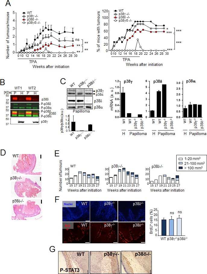
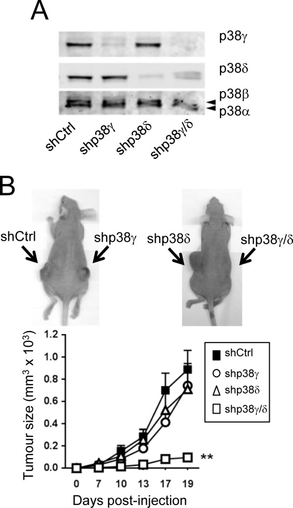
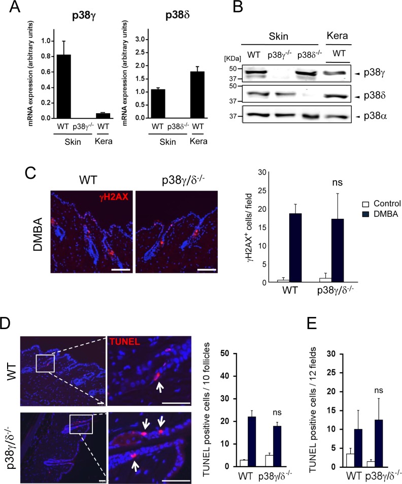
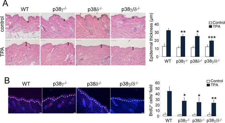
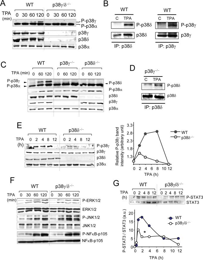
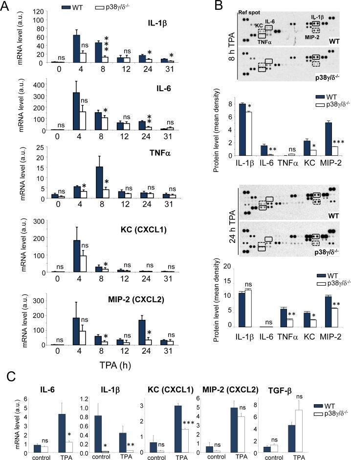
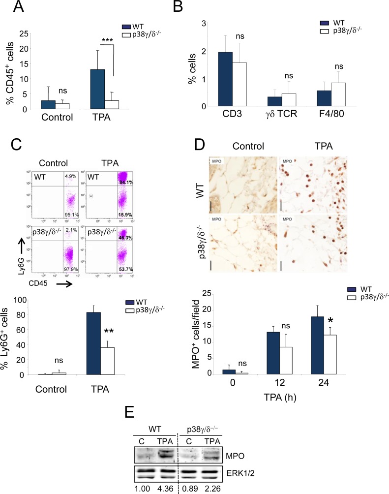
The contribution of chronic skin inflammation to the development of squamous cell carcinoma (SCC) is poorly understood. While the mitogen-activated protein kinase p38α regulates inflammatory responses and tumour development, little is known about the role of p38γ and p38δ in these processes. Here we show that combined p38γ and p38δ (p38γ/δ) deletion blocked skin tumour development in a chemically induced carcinogenesis model. p38γ/δ deletion reduced TPA-induced epidermal hyperproliferation and inflammation; it inhibited expression of proinflammatory cytokines and chemokines in keratinocytes in vitro and in whole skin in vivo, resulting in decreased neutrophil recruitment to skin. Our data indicate that p38γ/δ in keratinocytes promote carcinogenesis by enabling formation of a proinflammatory microenvironment that fosters epidermal hyperproliferation and tumourigenesis. These findings provide genetic evidence that p38γ and p38δ have essential roles in skin tumour development, and suggest that targeting inflammation through p38γ/δ offers a therapeutic strategy for SCC treatment and prevention.

Keywords: inflammation-associated cancer; knockout mice; p38γ; p38δ; skin.


Conflict of interest statement

CONFLICTS OF INTEREST

The authors declare that they have no conflict of interest.

Figures

Figure 1
Figure 1. p38γ/δ deletion reduces the incidence of DMBA/TPA-induced skin tumour formation in mice
A. Number of tumours per mouse and percentage of mice with tumours are shown at indicated times. WT (n = 9), p38γ−/− (n = 15), p38δ−/− (n = 16) and p38γ/δ−/− (n = 9) mice were treated with DMBA/TPA (see Methods) and the skin was monitored for tumour growth at indicated times. Tumour number per mouse shown as mean ± SEM. ns, not significant; *p ≤0.05; **p ≤ 0.01; ***p ≤ 0.001 relative to WT mice or between indicated genotypes (black lines). B. Papilloma (P) and healthy skin (H) protein extracts from two WT mice (WT1 and WT2) were immunoblotted with antibodies to active phosphorylated p38 (P-p38α, P-p38δ) (green), and total p38δ (red) and p38γ. Blots were analysed using the Odyssey infrared imaging system. P-p38δ was visualised in yellow when colours were merged. Results were similar in three independent experiments. C. Expression of p38γ, p38δ and p38α protein and mRNA. WT, p38γ−/− and p38δ−/− papilloma protein extracts were immunoblotted with the indicated antibodies. Representative blots are shown. Band intensities from the p38γ and p38α immunoblot were quantified using the Odyssey infrared imaging system. Quantification is represented as p38γ/p38α. Data show mean ± SEM. qPCR of p38MAPK mRNA in total RNA from WT, p38γ−/− or p38δ−/− papilloma and in total RNA from healthy skin (H) from WT mice. Expression of the different p38 mRNA was normalised to GAPDH. Data show mean ± SEM from one representative experiment of at least three with similar results. D. Representative H&E-stained sections of skin tumour at week 29 (Panel A). Scale bars: 500 μm. E. Histograms of tumour size distribution in A. at indicated times. F. Proliferation in tumours from WT, p38γ−/− and p38δ−/− mice (at week 29) was evaluated by BrdU staining. BrdU positive cells (red) were counted and represented as percentage of total basal keratinocytes. Nuclei are Hoechst33342-stained (blue). Results show mean ± SEM (n = 3-6 tumours/group). ns, not significant. Scale bars: 100 μm. G. Papilloma sections were stained to evaluate P-STAT3. Scale bars: 50 μm. Representative sections are shown. (See Materials and Methods).
Figure 2
Figure 2. Combined p38γ/δ deletion reduces tumourigenesis of A431 cells
A. shControl-, shp38γ-, shp38δ- and shp38γ/δ-A431 cell extracts (50 μg) were examined by immunoblotting with the indicated antibodies to determine p38γ and p38δ expression. B. Immunodeficient nude mice received subcutaneous injections of shControl-A431 cells, shp38γ-A431 cells, shp38δ-A431 cells and shp38γ/δ-A431 cells, and tumour volume was measured periodically as indicated. Values are means ± SD for 12 mice. Inset: Representative photographs of mouse tumour at day 19. Arrows indicate injection sites.
Figure 3
Figure 3. Combined p38γ and p38δ deletion does not affect DMBA response in the skin
A. p38γ and p38δ expression in the skin and in keratinocytes. qPCR of p38MAPK mRNA in total RNA from WT, p38γ−/− or p38δ−/− skin and in total RNA from keratinocytes from WT mice. Expression of the different p38 mRNA was normalised to GAPDH. Data show mean ± SEM (n = 3 mice/group). B. WT, p38γ−/− and p38δ−/− skin extracts and WT keratinocytes lysates (50 μg) were immunoblotted with antibodies to total p38γ, p38δ and p38α. Representative blots are shown. C. WT and p38γ/δ−/− mice were treated for 24 h with DMBA or acetone as control. Skin sections were immunofluorescence-stained to evaluate DMBA-induced DNA damage response (γH2AX). γH2AX+ cells (red) were quantified; at least 8 fields/mouse were scored. Results show mean ± SEM (n = 4 mice/group). ns, not significant, relative to WT mice in the same conditions. Scale bars: 100 μm. D., E. Apoptosis in mouse skin was evaluated by TUNEL staining (red) at 24 h post-DMBA application. Apoptotic cells were counted; 12 fields/mouse were scored. Results show mean ± SEM (n = 4 mice/group). ns, not significant, relative to WT mice. Panel D. shows quantitation of follicular apoptotic cells and E. shows interfollicular apoptotic cells. Scale bars: 50 μm. In C. and D. nuclei are Hoechst33342-stained (blue).
Figure 4
Figure 4. p38γ and p38δ deletion decreases epithelial cell proliferation
A. Representative H&E staining of skin sections from WT, p38γ−/−, p38δ−/− and p38γ/δ−/− mice treated for 31 h with TPA or acetone as control. Epidermal thickness, indicated by black lines, was measured. Results show mean ± SEM (n = 4 mice/group), *p ≤ 0.05; **p ≤ 0.01; ***p < 0.001. Scale bars: 100 μm. B. Proliferation in skin of WT, p38γ−/−, p38δ−/− and p38γ/δ−/− mice was evaluated by BrdU staining at 31 h post-TPA application. BrdU positive cells (red) were counted; 12 fields/mouse were usually scored. Nuclei are Hoechst33342-stained (blue). Results show mean ± SEM (n = 4 mice/group). *p ≤ 0.05; **p < 0.01, relative to WT mice. Scale bars: 50 μm.
Figure 5
Figure 5. Activation of intracellular signalling pathways by TPA
A. Skin extracts (50 μg) from WT and p38γ/δ−/− mice, treated with acetone (control, time 0) or with TPA for the indicated times, were immunoblotted with antibodies to active phosphorylated p38 (P-p38α, P-p38γ) and total p38α, p38γ and p38δ. B. WT mice were treated with TPA for 120 min as in A.. Endogenous p38δ and p38γ were immunoprecipitated from WT skin extracts (2 mg). Pellets were immunoblotted with anti-P-p38 (P-p38δ, P-p38γ) or -p38δ and -p38γ antibodies. Representative blots are shown. C. Skin protein extracts (50 μg) from WT, p38γ−/− and p38δ−/− treated with TPA for the indicated times, were immunoblotted with antibodies to active phosphorylated p38 (P-p38α, P-p38γ), and total p38α, p38γ and p38δ. Results were similar in three independent experiments. D. p38γ−/− mice were treated with TPA for 120 min as in C.. Endogenous p38δ was immunoprecipitated from skin extracts (2 mg). Pellets were immunoblotted with anti-P-p38 (P-p38δ) or -p38δ antibodies. Representative blots are shown. E. Skin protein extracts (50 μg) from WT and p38δ−/− mice, treated with TPA for the indicated times, were immunoblotted with antibodies as in C.. Bands from the immunoblots were quantified using the Odyssey infrared imaging system. Quantification is represented as P-p38γ/p38γ. Data show mean ± SEM. F. WT and p38γ/δ−/− mouse skin were treated as in A.. Skin extracts (50 μg) were immunoblotted with antibodies to active phosphorylated ERK1/2 (P-ERK1/2), active phosphorylated JNK1/2 (P-JNK1/2) or phosphorylated NF-κB-p105. Total protein levels of ERK1/2, JNK1/2, and NF-κB-p105 were also measured in the same lysates as loading controls. G. Skin extracts (50 μg) from control and TPA-treated WT and p38γ/δ−/− mice were immunoblotted with antibodies to phospho- and total STAT3. Representative blots are shown. Bands were quantified using the Odyssey infrared imaging system. Quantification is represented as densities ratio P-STAT3 /STAT3. *p ≤0.05.
Figure 6
Figure 6. p38γ/δ deletion reduces TPA-induced cytokine and chemokine production in mouse skin
A. Relative mRNA expression of indicated genes at different times was determined by qPCR in TPA-treated WT and p38γ/δ−/− mouse skin and normalised to GAPDH mRNA. Data show mean ± SEM (n = 3-6). ns, not significant; *p ≤ 0.05; **p ≤ 0.01, ***p ≤ 0.001, relative to WT mice in the same conditions. B. Skin protein extracts from three WT and three p38γ/δ−/− mice (300 μg total), treated with TPA for 8 (top) and 24 h (bottom), were mixed with an antibody mixture and incubated with the Mouse Cytokine Array Panel A membrane as indicated by the manufacturer (R&D Systems). Pixel densities on the film were analysed using ImageJ software. ns, not significant; *p ≤ 0.05; **p ≤ 0.01, ***p ≤ 0.001. C. Relative mRNA expression was determined by qPCR for indicated genes in TPA-treated WT and p38γ/δ−/− keratinocytes and normalised to GAPDH mRNA. In panel B. and C. data show mean ± SEM (n = 3). ns, not significant; *p ≤ 0.05; **p ≤ 0.01, ***p ≤ 0.001, relative to WT in the same conditions.
Figure 7
Figure 7. Reduced neutrophil recruitment in p38γ/δ−/− mice
A.-C. Skin cells from 24-h TPA-treated (or acetone as control) WT and p38γ/δ−/− mice were stained with anti-CD45, -CD3, -γδ TCR, -F4/80 and -Ly6G antibodies. A. Percentages of CD45+ cells are shown. CD45+ cells were gated and percentages of B. CD3+, γδ TCR+ and F4/80+ cells, or C. Ly6G+ cells were analysed by flow cytometry. Data show mean ± SEM (n = 3-4 per experiment and condition); **p ≤ 0.01, ***p ≤ 0.001, ns, not significant. Representative profiles are shown in C.. (D, E) WT or p38γ/δ−/− mice were treated for 12 or 24 h D. and for 24 h E. with TPA or acetone as control. D. Skin sections were immunohistochemically stained to evaluate neutrophils (MPO). MPO+ cells were quantified; 30 fields/mouse were usually scored. Results show mean ± SEM (n = 3 mice/group). *p ≤ 0.05; ns, not significant; Scale bars: 20 μm. E. WT and p38γ/δ−/− skin lysates (50 μg) were immunoblotted with antibodies to MPO and ERK1/2 (loading control). Immunoblots were quantified using the Odyssey infrared imaging system; normalised MPO band densities are represented numerically below the blot.

Similar articles

Cited by

References

    1. Brantsch K.D., Meisner C., Schonfisch B., Trilling B., Wehner-Caroli J., Rocken M., Breuninger H. Analysis of risk factors determining prognosis of cutaneous squamous-cell carcinoma: a prospective study. Lancet Oncol. 2008;9:713–720. - PubMed
    1. Garcia-Zuazaga J., Olbricht S.M. Cutaneous squamous cell carcinoma. Adv Dermatol. 2008;24:33–57. - PubMed
    1. Hanahan D., Coussens L.M. Accessories to the crime: functions of cells recruited to the tumor microenvironment. Cancer Cell. 2012;21:309–322. - PubMed
    1. Baldursson B., Sigurgeirsson B., Lindelof B. Leg ulcers and squamous cell carcinoma. An epidemiological study and a review of the literature. Acta Derm Venereol. 1993;73:171–174. - PubMed
    1. Motswaledi M.H., Doman C. Lupus vulgaris with squamous cell carcinoma. J Cutan Pathol. 2007;34:939–941. - PubMed

Publication types

MeSH terms

Substances