. 2015 Aug 21;6(24):20697-710.
doi: 10.18632/oncotarget.4136.

AF1q is a novel TCF7 co-factor which activates CD44 and promotes breast cancer metastasis

Affiliations

AF1q is a novel TCF7 co-factor which activates CD44 and promotes breast cancer metastasis

Jino Park et al. Oncotarget. .

Abstract

AF1q is an MLL fusion partner that was identified from acute myeloid leukemia (AML) patients with t (1; 11) (q21; q23) chromosomal abnormality. The function of AF1q is not yet fully known, however, elevated AF1q expression is associated with poor clinical outcomes in various malignancies. Here, we show that AF1q specifically binds to T-cell-factor-7 (TCF7) in the Wnt signaling pathway and results in transcriptional activation of CD44 as well as multiple downstream targets of the TCF7/LEF1. In addition, enhanced AF1q expression promotes breast cancer cell proliferation, migration, mammosphere formation, and chemo-resistance. In xenograft models, enforced AF1q expression in breast cancer cells also promotes liver metastasis and lung colonization. In a cohort of 63 breast cancer patients, higher percentages of AF1q-positive cancer cells in primary sites were associated with significantly poorer overall survival (OS), disease-free survival (DFS), and brain metastasis-free survival (b-MFS). Using paired primary/metastatic samples from the same patients, we demonstrate that AF1q-positive breast cancer cells become dynamically dominant in the metastatic sites compared to the primary sites. Our findings indicate that breast cancer cells with a hyperactive AF1q/TCF7/CD44 regulatory axis in the primary sites may represent "metastatic founder cells" which have invasive properties.

Keywords: AF1q; CD44; TCF7; Wnt; metastasis.


Conflict of interest statement

CONFLICTS OF INTEREST

The authors disclose no potential conflict of interest.

Figures

Figure 1
Figure 1. Effects of AF1q expression in breast normal epithelial (HMLE and MCF10a) and cancer cell lines (MDA-MB-231 and MDA-MB-231LN)
A. Immunohistochemistry (IHC) staining shows a higher frequency and intensity of AF1q-positive cancer cells in metastatic sites compared to primary sites. In normal breast tissue, AF1q staining was restricted to myoepithelial cells rather than ductal/glandular epithelial cells. Scale bars = 50 μm. B. Enforced AF1q expression in breast normal epithelial and cancer cell lines promoted transwell migration and matrigel invasion compared to their correspondent empty vector (EV) controls. Using short hairpin RNA (shRNA) to suppress the endogenous AF1q expression in MCF10a and MDA-MB-231LN inhibited transwell migration and matrigel invasion compared to their correspondent scrambled RNA (Scr) controls. C. Enforced AF1q expression promoted mammosphere formation in breast normal epithelial cell lines (HMLE and MCF10a) and tumorsphere formation in breast cancer cell lines (MDA-MB-231 and MDA-MB-231LN); suppression of the endogenous AF1q expression with shRNA in MCF10a or MDA-MB-231LN inhibited mammosphere or tumorsphere formation, respectively. D. Endogenous AF1q expression in MDA-MB-231LN was significantly elevated after treatment for 24 hours with two different concentrations of doxorubicin or etoposide compared to its controls. Cell viability was significantly decreased with 100 nM doxorubicin and 100 uM etoposide (lower than 50%), but not with lower concentration of both. Data are mean values ± s.d. of three independent experiments. P values were calculated using student t test (*, P < 0.05; **, P < 0.01; ***, P < 0.001).
Figure 2
Figure 2. AF1q is a cofactor of TCF7 and forms a transcriptional complex with TCF7, LEF1 and β-catenin which leads to a transcriptional activation of CD44, one of the downstream targets of the Wnt signaling pathway
A. AF1q and TCF7 interaction was confirmed in MCF10a by co-immunoprecipitation of TCF7 with anti-AF1q antibody. NC is negative control for nonspecific binding. B. The Schrodinger program generates a three-dimensional AF1q and TCF7 interaction model based on yeast 2-Hybrid results; TCF7 forms a glove-like shape to envelope the entire AF1q protein. C. The AF1q sequence is highly conserved across mammalian species, in particular the acidic (E and D in red) and phosphorylatable amino acids (S, T and Y in green) are highlighted. The acidic nature is best reflected by the low isoelectric point (pI) of the peptides across different species. There is a repeated internal protein motif region (aa 8-30 and aa 68-90) in the 9 kDa peptide of AF1q shown below the AF1q amino acid sequences between different mammalian species. D. TOP/FLOP flash reporter assays show that enforced AF1q expression enhanced the Wnt signaling activity; the suppressed endogenous AF1q expression by shRNA attenuated the Wnt signaling activity. E. AF1q enhanced CD44 promoter activity, as shown by comparing transfection of the pGL4-CD44 promoter with the pGL4-EV reporter control. F. Super-shift assay using the MCF10a nuclear extracts showed that AF1q and TCF7 directly bind to the CD44 promoter. G. The AF1q nuclear shift after enforced AF1q expression was confirmed in fractionated cytoplasm and nuclear extracts from MCF10a cells. Data are mean values ± s.d. of three independent experiments. P values were calculated using student t test (*, P < 0.05; **, P < 0.01; ***, P < 0.001).
Figure 3
Figure 3. AF1q mediated cell transwell migration, matrigel invasion, and mammosphere (tumor sphere) formation essentially depends on activation of CD44
A. CD44 expression was up-regulated after enforced AF1q expression in all tested cell lines. CD44 expression was attenuated after suppressed endogenous AF1q expression by shRNA in MCF10a and MDA-MB-231LN. B. Suppressed CD44 expression by shRNA in MCF10a/AF1q and MDA-MB-231LN/AF1q did not affect AF1q expression. C. Suppressed CD44 expression by shRNA in MCF10a/AF1q and MDA-MB-231LN/AF1q attenuated AF1q-mediated transwell migration and matrigel invasion. D. Suppressed CD44 expression by shRNA in MCF10a/AF1q and MDA-MB-231LN/AF1q attenuated AF1q-mediated mammosphere/tumor sphere formation. Data are mean values ± s.d. of three independent experiments. P values were calculated using student t test (*, P < 0.05; **, P < 0.01; ***, P < 0.001).
Figure 4
Figure 4. AF1q is tightly associated with a metastasis gene cluster
A. RNA-seq analysis suggests that enforced or suppressed AF1q expression by lentiviral system led to up-regulation or down-regulation of 243 and 120 genes, respectively. Pathway activities significantly affected by enforced AF1q expression are illustrated in bar graph (www.ingenuity.com; black squares indicate P-value). B. Hierarchical clustering of AF1q associated multiple downstream targets in the Wnt signaling pathway are depicted in a Heat map. The dendrogram groups genes into distinct groups according to their distance from each other. The distance measure is shown on the horizontal axis. Results are from MDA-MB-231LN with enforced AF1q expression. Down-regulated genes are shown in green and up-regulated genes in red. Fold change corresponds to AF1q over Ctrl-transduction. The table shows FDR-adjusted p-value (‘q-value) as well as the uncorrected p-value of the test statistic.
Figure 5
Figure 5. NOD/SCID xenograft mouse models confirm the association of AF1q with breast cancer metastasis
A. Representative image of mice injected with MDA-MB-231LN Con, AF1q, Scr and shRNA cells (upper panel) and tumor, liver, lung, kidney and spleen tissues dissected from the 4 treated groups of mice (lower panel) at 4 weeks after injection in mammary fat pad model. B. Tumors were harvested from mammary fat pad injection model and size and weight were measured at 4 weeks after injection. MDA-MB-231LN/AF1q injected mice grew significantly larger tumor masses compared to MDA-MB-231LN/EV control mice. Suppressed the endogenous AF1q expression with shRNA in MDA-MB-231LN were significantly smaller and lighter tumors. The tumor weight is shown as the mean ± s.d. P values were calculated using student t test (*, P < 0.05; **, P < 0.01; ***, P < 0.001). C. The tail vein injection model, MDA-MB-231LN/AF1q significantly grew larger tumor masses than MDA-MB-231LN/EV. Data were mean values ± s.d. P values were calculated using student t test (*, P < 0.05; **, P < 0.01; ***, P < 0.001). D. Representative histology sections from the tail vein injection model MDA-MB-231LN/AF1q displayed more aggressive histologic features than controls (upper panel); arrows indicate areas of metastasis formation. Obvious liver metastases developed in both mammary pad (2/3 mice) and tail vein injection (1/3 mice) models (lower panel), but not in controls. Scale bars = 50 μm.
Figure 6
Figure 6. AF1q expression in breast cancer is positively associated with metastasis
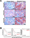
A. Representative sections of paired primary/metastatic tissue from 3 breast cancer patients demonstrated a significantly higher frequency and intensity of AF1q-positive breast cancer cells in metastatic sites (lymph nodes and lung) than in primary sites. Scale bars = 50 μm. B. Breast cancer patients with ≥50% AF1q-positive cancer cells in primary diagnostic sites (high, n = 21) had significantly poorer overall survival (OS), disease-free survival (DFS) and brain metastasis-free survival (b-MFS) than patients with < 50% AF1q-positive cancer cells (Low, n = 42).

References

    1. Tse W, Zhu W, Chen HS, Cohen A. A novel gene, AF1q, fused to MLL in t(1;11) (q21;q23), is specifically expressed in leukemic and immature hematopoietic cells. Blood. 1995;85:650–656. - PubMed
    1. Xiong Y, Li Z, Ji M, Tan AC, Bemis J, Tse JV, Huang G, Park J, Ji C, Chen J, Bemis LT, Bunting KD, Tse W. MIR29B regulates expression of MLLT11 (AF1Q), an MLL fusion partner, and low MIR29B expression associates with adverse cytogenetics and poor overall survival in AML. British journal of haematology. 2011;153:753–757. - PMC - PubMed
    1. Tiberio P, Cavadini E, Callari M, Daidone MG, Appierto V. AF1q: a novel mediator of basal and 4-HPR-induced apoptosis in ovarian cancer cells. PloS one. 2012;7:e39968. - PMC - PubMed
    1. Co NN, Tsang WP, Tsang TY, Yeung CL, Yau PL, Kong SK, Kwok TT. AF1q enhancement of gamma irradiation-induced apoptosis by up-regulation of BAD expression via NF-kappaB in human squamous carcinoma A431 cells. Oncology reports. 2010;24:547–554. - PubMed
    1. Jacques C, Baris O, Prunier-Mirebeau D, Savagner F, Rodien P, Rohmer V, Franc B, Guyetant S, Malthiery Y, Reynier P. Two-step differential expression analysis reveals a new set of genes involved in thyroid oncocytic tumors. The Journal of clinical endocrinology and metabolism. 2005;90:2314–2320. - PubMed

Publication types

MeSH terms