. 2015 Jun 30;112(26):E3365-73.
doi: 10.1073/pnas.1502497112. Epub 2015 Jun 15.

Self-repairing symmetry in jellyfish through mechanically driven reorganization

Affiliations

Self-repairing symmetry in jellyfish through mechanically driven reorganization

Michael J Abrams et al. Proc Natl Acad Sci U S A. .

Abstract

What happens when an animal is injured and loses important structures? Some animals simply heal the wound, whereas others are able to regenerate lost parts. In this study, we report a previously unidentified strategy of self-repair, where moon jellyfish respond to injuries by reorganizing existing parts, and rebuilding essential body symmetry, without regenerating what is lost. Specifically, in response to arm amputation, the young jellyfish of Aurelia aurita rearrange their remaining arms, recenter their manubria, and rebuild their muscular networks, all completed within 12 hours to 4 days. We call this process symmetrization. We find that symmetrization is not driven by external cues, cell proliferation, cell death, and proceeded even when foreign arms were grafted on. Instead, we find that forces generated by the muscular network are essential. Inhibiting pulsation using muscle relaxants completely, and reversibly, blocked symmetrization. Furthermore, we observed that decreasing pulse frequency using muscle relaxants slowed symmetrization, whereas increasing pulse frequency by lowering the magnesium concentration in seawater accelerated symmetrization. A mathematical model that describes the compressive forces from the muscle contraction, within the context of the elastic response from the mesoglea and the ephyra geometry, can recapitulate the recovery of global symmetry. Thus, self-repair in Aurelia proceeds through the reorganization of existing parts, and is driven by forces generated by its own propulsion machinery. We find evidence for symmetrization across species of jellyfish (Chrysaora pacifica, Mastigias sp., and Cotylorhiza tuberculata).

Keywords: jellyfish; propulsion; reorganization; self-repair; symmetry.


Conflict of interest statement

The authors declare no conflict of interest.

Figures

Fig. 1.
Fig. 1.
Life cycle and anatomy of Aurelia aurita. (A) Adult Aurelia. The blue color is due to lighting. Image courtesy of Wikimedia Commons/Hans Hillewaert. Image © Hans Hillewaert. (B) Aurelia life cycle. Fertilized eggs develop into larval planulae, which settle and develop into polyps. Seasonally, or in the right conditions, the polyps metamorphose into strobilae and release free-swimming, juvenile jellyfish (a process called strobilation). The young jellyfish, called ephyrae, grow into medusae in 3–4 wk. Reprinted with permission from ref. . (C) A juvenile green sea turtle preying on Aurelia at Playa Tamarindo, Puerto Rico. Image courtesy of R. P. van Dam. (D) An Aurelia ephyra has eight radially symmetrical arms, surrounding the manubrium at the center. At the end of each arm is a light- and gravity-sensing organ, called rhopalium. (E) The epithelium of ephyra is composed of two cell layers, the ectoderm-derived epidermis that faces the outer side and the endoderm-derived gastrodermis that lines the gastric cavity. Between the two layers is the gelatinous, viscoelastic mesoglea. Embedded in the subumbrellar side (mouth side) is the coronal muscle (green).
Fig. 2.
Fig. 2.
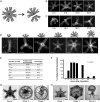
Aurelia ephyra reorganize existing arms to regain radial symmetry. (A) An example of amputation schemes used in the study. Cuts were performed across the body using a razor blade. (B and C) A three-armed and five-armed piece amputated from a single ephyra. Within 2 d, neither regenerated the lost arms. Instead, each reorganized to reform radial symmetry. (D) Symmetrization was observed with two, three, four, five, six, and seven arms. The cartoons indicate the initial forms after amputation. (E) Percentage of symmetrization across amputation schemes. The ephyrae in the amputation experiments were 1–3 d old (after strobilation) and were examined daily for 4 d. (F) Progression of symmetrization. In this experiment, we counted the number of ephyrae that symmetrized at the indicated time. Data were collected from dimers, tetramers, pentamers, and hexamers. There is a slight trend in the recovery speed across amputation scheme. The 12-h recovery is typical for dimers. Symmetrical tetramers and pentamers often started appearing by day 1 onward, as analyzed in more detail in Fig. 5H. (G and H) Ephyrae in these experiments were tracked individually for 1 mo, fed daily, and imaged every 2–3 d. (G) Pentamers that symmetrized continued growing into mature medusa (n = 19). (H) Pentamers that did not symmetrize grew abnormally with oversized manubria (n = 10). (Scale bar in each photograph: 1 mm.)
Fig. S1.
Fig. S1.
Symmetrization proceeded with foreign arms. (A) In this experiment, the ephyra was cut in half. Subsequently, an arm from another ephyra (outlined in red) was grafted to the tetramer, between arm 1 and 2. “m” indicates the manubrium. Grafting was performed by pinning the ephyra segments next to each other on an agarose plate (1% agarose, made with artificial seawater). Pinning was done using cactus spines (ones from columnar Espostoa sp. worked best). The ephyrae were kept pinned overnight (∼12 h), unpinned the next morning, and allowed to recover in artificial seawater. (B) By 4 d, the patchwork ephyra had become symmetrical. The grafted arm is outlined in red. The location of grafting looked smooth, and the ephyra had healed without obvious scarring. The extra arm was incorporated seamlessly into the host tetramer. The resulting patchwork pentamer was symmetrical and pulsed synchronously. (C) Phalloidin staining shows that the axisymmetrical muscle was rebuilt, and muscle from the extra arm was connected seamlessly into the host ephyra (we will discuss the muscle network in more detail in the main text and in Fig. 4).
Fig. 3.
Fig. 3.
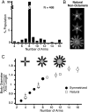
Symmetrization phenocopies developmental variation. (A) Nonoctamers form 9.5% of the Aurelia population in our laboratory. Ephyrae were scored immediately upon strobilation. This histogram come from multiple strobilae in a single strobilation round. A single strobila may produce 10–20 ephyrae, with variable numbers of arms. (B) A natural pentamer, hexamer, and dodecamer. (C) White circles: body size of natural ephyrae. Black circles: body size of ephyrae from symmetrization. Both plotted as a function of the arm number. The arrows indicate where there are both black and white circles overlapping. Body size was measured as the diameter (the gray region in the ephyra cartoons). We normalized body diameter to arm length (black regions of the ephyra cartoons), to account for variation across ephyrae. The ephyrae also grew in size over time; to account for this, we characterized the growth curve and normalized all measurements to 1-d-old ephyrae (Materials and Methods). A total of 46 ephyrae was measured to generate this plot. Error bars are SD from more than three ephyrae. Some error bars are not seen because they are smaller than the circles.
Fig. 4.
Fig. 4.
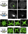
Symmetrization is not driven by cell proliferation, cell death, or muscle reconnection. (A–D) Is symmetrization driven by cell proliferation? (A) Localized cell proliferation (e.g., in the green regions) may push the arms apart. (B) EdU stain (green) in a symmetrized tetramer, showing cumulative signal over 4 d. (C) EdU stain was abolished in the presence of 20 μM hydroxyurea. In this experiment, the cut ephyrae were incubated in EdU with or without 20 μM hydroxyurea for 4 d. The solution was refreshed daily. Ephyrae were fixed and stained on day 4 (Materials and Methods). (D-F) Is symmetrization driven by cell death? (D) Localized cell death (e.g., in the blue region) may pull the arms into the cut site. (E) Sytox stain (white) in a symmetrized ephyra 3 d after amputation. (F) Sytox stain was abolished in the presence of a caspase inhibitor (100 μM Z-vad-fmk). Cut ephyrae were incubated in the inhibitor for 3 d, and then stained with Sytox (Materials and Methods). (G–J) Symmetrization is accompanied by reconnection of coronal muscle. (G) Staining of the musculature in an uncut ephyra. Muscle was visualized using phalloidin–Alexa Fluor 488 (Materials and Methods). (H–J) Staining of muscle in symmetrizing ephyrae. Ephyrae were fixed and stained at 15 min (H), 1 d (I), and 3 d after amputation (J). White arrows in K indicate the extending edges of the muscle. (K–N) Is symmetrization driven by muscle reconnection? (K) Reconnection of muscle (green) may pull the arms along. (L–N) Ephyrae were amputated, incubated in 2 μM (L–M) or 500 nM (N) cytochalasin D for 4 d, and then stained with phalloidin–Alexa Fluor 488.
Fig. S2.
Fig. S2.
Cell proliferation and cell death stains. (A) EdU stain (green) in an uncut ephyrae. Total nuclei were stained using Hoechst (white). The magnified regions show the EdU and nuclear stain separately. The circle indicates the manubrium. (B) Sparse baseline Sytox stain (white) in an uncut ephyra. (C) Sytox stain was increased in the presence a caspase inducer (100 nM gambogic acid; n = 19 of 20). In this experiment, cut ephyrae were incubated in the chemicals for 1–3 d, and then stained with Sytox (Materials and Methods). The 1 μM gambogic acid was lethal to ephyrae, giving us an upper limit.
Fig. 5.
Fig. 5.
Symmetrization is driven by muscle contraction in the propulsion machinery. (A–C) Inhibiting muscle contraction blocks symmetrization. (A) Amputated ephyrae were incubated in 400 μM menthol for 4 d, and then stained with phalloidin. All treated ephyrae failed to symmetrize (n = 60 of 60). (B) A magnified view shows that the cut muscle remained blunt in the presence of menthol. (C) Ephyrae removed from menthol (after 4 d) resumed and completed symmetrization within 4 d (n = 20 of 20). (D and E) Proposed model of symmetrization. (D) A swimming stroke consists of muscle contraction, which generates compression, followed by elastic response from the mesoglea. We propose that, in the amputated ephyrae, this leads to angular pivoting into the cut site, as there is less bulk resistance. With repeated cycles of compression and elastic repulsion, the arms gradually relax into a more symmetrical state, until the forces are rebalanced. (E) Mathematical simulation of the symmetrization of a tetramer, taking into account the compression generated by the muscle contraction, the elastic response, and the ephyra geometry (see Supporting Information for details of the model). The predicted time of symmetrization is computed based on the pulsation frequency measured in seawater (Fig. 5F). (F–H) Frequency of muscle contraction dictates the speed of symmetrization. (F) Incubation in reduced MgCl2 (50% of the normal seawater) increased the frequency of muscle contraction, whereas incubation in 80 μM menthol decreased the frequency of muscle contraction. The dashed gray line shows the full range of the data, whereas the black lines indicate 95% confidence intervals. (G) Sample traces of ephyra pulsation in normal seawater (blue), reduced magnesium (black), and 80 μM menthol (green). Frequency of muscle contraction was counted by hand from time-lapse movies taken at 15 fps. A single pulse typically takes 0.5 s. Full contraction was when the ephyrae fully closed in, and partial contraction was when the arms only contracted halfway. (H) Cut ephyrae were incubated in normal seawater (blue), seawater with reduced MgCl2 (black), or 80 μM menthol (green), and scored every day for symmetrization.
Fig. S3.
Fig. S3.
Inhibitors of skeletal myosin II blocks symmetrization. In this experiment, the ephyrae were cut, and then incubated in (A) 2,3-butanedione monoxime (BDM) (25 mM) or (B) N-benzyl-p-toluene-sulfonamide (BTS) (150 mM). In both inhibitors, the ephyrae did not pulse and remained asymmetrical throughout the 4-d treatment, and the coronal muscle remained blunt (n = 40 of 40 for BDM; n = 40 of 40 for BTS). The phalloidin staining was performed on day 4 after amputation.
Fig. S4.
Fig. S4.
Symmetrization was observed across four species of Scyphozoan jellyfish. (A) The moon jellyfish Aurelia aurita. Image courtesy of Wikimedia Commons/Hans Hillewaert. Image © Hans Hillewaert. (B) The sea nettle Chrysaora pacifica. Image courtesy of Sofi Quinodoz. (C) The lagoon jellyfish Mastigias sp. Image courtesy of Wikimedia Commons/Captmondo. (D) The Mediterranean jellyfish Cotylorhiza tuberculata. Image courtesy of Wikimedia Commons/Antonio Sontuoso. For each column, row 1 shows the adult medusa, row 2 shows the uncut ephyra, and row 3 shows the symmetrized tetramer from amputation. Freshly strobilated ephyrae were cut in half and allowed to recover in seawater. Symmetrized tetramers were observed within 4 d in all species.
Fig. S5.
Fig. S5.
Removing the manubrium broke planarity. (A) Punching out the manubrium led the ephyrae to adopt a fan shape. We used a P200 pipette tip to make a clean hole and removed the entire manubrium. (B) Punching out the manubrium and linearizing the ribbon led the ephyrae to adopt a spiral shape, with threefold symmetry stacked on fivefold symmetry. The new symmetry was observed within 1–4 d after amputation. The ephyrae pulsed synchronously and remained alive for over 3 wk until the experiment was ended.
Fig. S6.
Fig. S6.
Model geometry and coordinate. (A) We consider a Δθi, the angular span between arm i and arm i + 1. (B) The corresponding body area is denoted as Ai. Muscle contraction compresses the body by ΔA (C) and generates an elastic response (D), which in turn leads to angular pivoting of the arms (E).
Fig. 6.
Fig. 6.
(A) Line indicates arm length. Red area indicates the body. (B) Body size increases linearly with age. Error bars were from more than three biological replicates and technical replicates.

References

    1. Graham WM, Kroutil RM. Size-based prey selectivity and dietary shifts in the jellyfish, Aurelia aurita. J Plankton Res. 2001;23(1):67–74.
    1. Purcell JEC, Graham WMC, Dumont HJC. 2001. Jellyfish Blooms: Ecological and Societal Importance. Proceedings of the International Conference on Jellyfish Blooms, held in Gulf Shores, Alabama, 12–14 January, 2000. Hydrobiologia (Springer Science and Business Media, Dordrecht, The Netherlands), Vol 451, p 2001.
    1. Olesen NJ, Riisgård HU. Population dynamic, growth and energetics of jellyfish, Aurelia aurita, in a shallow fjord. Mar Ecol Prog Ser. 1994;105:9–18.
    1. Lucas CH. Reproduction and life history strategies of the common jellyfish, Aurelia aurita, in relation to its ambient environment. Hydrobiologia. 2001;451(1-3):229–246.
    1. Conley K, Uye SI. Effects of hyposalinity on survival and settlement of moon jellyfish (Aurelia aurita) planulae. J Exp Mar Biol Ecol. 2015;462:14–19.

Publication types

LinkOut - more resources