. 2015 Jun 30;112(26):E3327-36.
doi: 10.1073/pnas.1509658112. Epub 2015 Jun 16.

Arginine methylation of HSP70 regulates retinoid acid-mediated RARβ2 gene activation

Affiliations

Arginine methylation of HSP70 regulates retinoid acid-mediated RARβ2 gene activation

Wei-wei Gao et al. Proc Natl Acad Sci U S A. .

Abstract

Although "histone" methyltransferases and demethylases are well established to regulate transcriptional programs and to use nonhistone proteins as substrates, their possible roles in regulation of heat-shock proteins in the nucleus have not been investigated. Here, we report that a highly conserved arginine residue, R469, in HSP70 (heat-shock protein of 70 kDa) proteins, an evolutionarily conserved protein family of ATP-dependent molecular chaperone, was monomethylated (me1), at least partially, by coactivator-associated arginine methyltransferase 1/protein arginine methyltransferase 4 (CARM1/PRMT4) and demethylated by jumonji-domain-containing 6 (JMJD6), both in vitro and in cultured cells. Functional studies revealed that HSP70 could directly regulate retinoid acid (RA)-induced retinoid acid receptor β2 (RARβ2) gene transcription through its binding to chromatin, with R469me1 being essential in this process. HSP70's function in gene transcriptional regulation appears to be distinct from its protein chaperon activity. R469me1 was shown to mediate the interaction between HSP70 and TFIIH, which involves in RNA polymerase II phosphorylation and thus transcriptional initiation. Our findings expand the repertoire of nonhistone substrates targeted by PRMT4 and JMJD6, and reveal a new function of HSP70 proteins in gene transcription at the chromatin level aside from its classic role in protein folding and quality control.

Keywords: arginine methylation; gene transcription; heat-shock proteins.


Conflict of interest statement

The authors declare no conflict of interest.

Figures
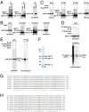
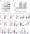
Fig. 1.
Fig. 1.
HSP70 methylation in cultured cells. (A) Cell lysates collected from HEK293T cells were subjected to IP with control IgG or anti-HSP70 antibody followed by IB with anti-methyl-lysine (Kme) (Left), antimethyl-arginine (Rme) (Center), or anti-HSP70 (Right) antibody. IgG H.C: IgG heavy chain. (B) HEK293T cells were treated with or without AdOx (20 μM) for 6 h before IP and IB experiments as described in A. (C) HEK293T cells were transfected with Flag-tagged HSP70 and then treated with or without AdOx (20 μM) for 6 h before IP with anti-Flag antibody, followed by IB with anti-Kme, anti-Rme, or anti-Flag antibody. Input was shown on the far left panel. N.S: nonspecific bands. (D) In vivo labeling of HSP70 proteins. HEK293T cells were maintained in DMEM without methionine for 24 h before adding NTM [cycloheximide (100 μg/mL) and chloramphenicol (40 μg/mL)] for another hour, followed by adding [35S]-methionine (20 μCi/mL) (lanes 1 and 2) or l-[methyl-3H]-methionine (20 μCi/mL) (lanes 3 and 4) in the presence or absence of AdOx (20 μM) for 4 h. Cells were then lysed and subjected to IP with anti-HSP70 antibody, followed by SDS/PAGE gel and autoradiogram. (E) HEK293T cells were transfected with Flag-tagged HSP70 before in vivo labeling with l-[methyl-3H]-methionine as described in D. (F) HSP70 proteins purified from HEK293T cells were subjected to Coomassie blue staining (C.B.S). IgG H.C: IgG heavy chain; IgG L.C: IgG light chain. (G and H) Sequence alignment of the region surrounding arginine 469 (boxed) in multiple HSP70 isoforms (Homo sapiens) (G) or in paralogous HSP70 genes in various organisms (H) by using Clustal Omega. Asterisk (*) indicates positions which have fully conserved residue; Colon (:) indicates conservation between groups of strongly similar properties; Period (.) indicates conservation between groups of weakly similar properties. At, Arabidopsis thaliana; Dm, Drosophila melanogaster; Dr, Danio rerio; Hs, Homo sapiens; Mm, Mus musculus; Pf, Plasmodium falciparum; Rn, Rattus norvegicus; Sc, Saccharomyces cerevisiae; Xt, Xenopus (Silurana) tropicalis.
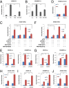
Fig. 2.
Fig. 2.
Methylation and demethylation of arginine 469 in HSP70 by PRMT4 and JMJD6, respectively. (A) Anti-HSP70R469me1 antibody was preincubated with 3XFlag (Left), HSP70R469 unmodified (Center), or HSP70R469me1 (Right) peptide for 30 min before probing HEK293T total cell lysates. (B) HEK293T cells transfected with control vector, Flag-tagged wild-type (WT) or mutant HSP70 (R469A) were subjected to IP with anti-Flag antibody, followed by IB with anti-HSP70R469me1 antibody. (C) HEK293T cells were transfected with control siRNA or siRNA specifically against each individual member of PRMT protein family for 72 h, followed by IB with anti-HSP70R469me1 (Upper) or anti-HSP70 (Lower) antibody. (D) In vitro methylation assay was performed by mixing purified bacterially expressed PRMTs with wild-type (Upper) or mutant HSP70 (R469A) (Lower) proteins. White arrows indicate automethylation of PRMT1, PRMT4, and PRMT6; Black arrows indicate methylation of HSP70. (E) In vitro methylation assay was performed by mixing purified bacterially expressed PRMT4 proteins with R469 or R469A peptide. Increased amount of each reaction was taken for dot blot as indicated and followed by autoradiogram. (F) In vitro demethylation assay was performed by mixing R469me1 peptide with or without purified bacterially expressed JMJD6 protein. Increased amount of each reaction was taken for dot blot as indicated and followed by IB with anti-HSP70R469me1 antibody. (G) HEK293T cells were transfected with control siRNA or siRNA specifically against JMJD6 for 72 h, followed by IB with antibodies as indicated. The ratio of R469me1 levels between the siCTL and siJMJD6 sample was 0.45 quantified using ImageJ.
Fig. S1.
Fig. S1.
Knock-down efficiency of siRNAs targeting PRMTs. Knock-down efficiency of siRNA targeting to each individual member of PRMT protein family as shown in Fig. 2C was examined through RT-qPCR. Of note, the expression level of PRMT8 was too low to be detected in HEK293T cells. Data shown is the relative fold-change, as indicated, compared with control siRNA transfected samples after normalization to actin (±SEM).
Fig. S2.
Fig. S2.
HSP70 methylation mediated by PRMTs. (A) In vitro purified, bacterially expressed His-tagged wild-type and mutant HSP70 (R469A) were examined by Coomassie blue staining (C.B.S). (B) In vitro purified, bacterially expressed GST-tagged PRMTs were examined by Coomassie blue staining. Of note, PRMT3 was purified separately. (C) Histones purified from HeLa cells (Sigma) were examined by Coomassie blue staining. (D) In vitro methylation assay was performed by mixing PRMTs with histones as described in B and C, respectively. Of note, in vitro methylation assay with PRMT3 was performed separately. (E) In vitro methylation assay was performed by mixing Flag-tagged PRMTs purified from overexpressed HEK293T cells with histones as described in C. (F) In vitro methylation assay was performed by mixing Flag-tagged PRMTs purified from overexpressed HEK293T cells with wild-type (Upper) or mutant HSP70 (R469A) (Lower) proteins. (G) The expression of Flag-tagged PRMTs in HEK293T cells was examined by IB with anti-Flag antibody. Of note, the expression of PRMT11 was low under current conditions tested. (H) In vitro methylation assay was performed by mixing PRMT1 protein as described in B, with R469 or R469A peptide. Increased amount of each reaction was taken for dot blot as indicated and followed by autoradiogram.
Fig. S3.
Fig. S3.
Demethylation of HSP70 R469me1 by JMJD6. In vitro demethylation assay was performed by mixing R469me1 peptide with (Lower) or without (Upper) purified bacterially expressed JMJD6 proteins, followed by MALDI-TOF MS analysis.
Fig. 3.
Fig. 3.
HSP70 association with chromatin in gene transcriptional regulation. (A) Cellular fractionation was done in three independent cell lines, HEK293T, HeLa, and MCF7, followed by IB with various antibodies, as indicated. (B) HSP70 ATPase activity assay was performed by mixing PRMT4 (0.04 μg/μL), HSP70 (wild-type or R469A mutant) (0.05 μg/μL), SAM (1 mM), and ATP (100 μM), as indicated. (C) HEK293T cells were treated with or without RA (10−7 M) for 1 h and then subjected to ChIP with control IgG, anti-HSP70, RXRα, or RARα antibody. Binding of HSP70, RXRα, and RARα on the RARβ2 gene promoter region were examined through qPCR. ChIP signals were presented as fold-enrichment over that of IgG (±SEM, **P < 0.01, ***P < 0.001). (D and E) MCF7 cells were treated with or without E2 (10−7 M) for 1 h and then subjected to ChIP with control IgG, anti-HSP70 (D) or ERα (E) antibody. Binding of HSP70 (D) and ERα (E) on TFF1/pS2 or GREB1 gene promoter regions were examined through qPCR (±SEM, *P < 0.05, **P < 0.01). (F and G) HEK293T (F) or MCF7 (G) cells were transfected with control siRNA or siRNA specifically targeting HSP70 for 72 h, followed by treatment with RA (10−7 M) (F) or E2 (10−7 M) (G), respectively, for 6 h before RT-qPCR analysis to examine mRNA levels of genes as indicated. Data shown was the relative fold-change compared with control siRNA transfected samples after normalization to actin (±SEM, *P < 0.05, **P < 0.01, ***P < 0.001). (H) Cell lysates as described in F and G were subjected to IB with antibodies, as indicated. (I) HEK293T cells were pretreated with or without PES (20 μM) or AMI (100 μM) for 1 h before treatment with or without RA (10−7 M) for 6 h, followed by RT-qPCR analysis to examine the mRNA levels of RARβ2. Data shown is the relative fold-change compared with control sample after normalization to actin (±SEM, ***P < 0.001).
Fig. S4.
Fig. S4.
Cellular localization of HSP70 and its R469-methylated form. (A) HEK293T cells were subjected to immunostaining with anti-HSP70 antibody. HSP70 proteins are shown in green. (B) Cellular fractionation was done in three independent cell lines, HCT116, SW480, and LNCaP, followed by IB with various antibodies as indicated.
Fig. S5.
Fig. S5.
Transcriptional regulation by HSP70. (A) NTera2 cells were treated with or without RA (10−7 M) for 1 h and then subjected to ChIP with control IgG or anti-HSP70 antibody. Binding of HSP70 on selected Hox gene promoter regions as indicated was examined through qPCR. ChIP signals were presented as fold-enrichment over that of IgG (±SEM, **P < 0.01, ***P < 0.001). (B) NTera2 cells were infected with control shRNA or shRNA specifically targeting HSP70 lentiviral particles for 72 h, followed by treatment with or without RA (10−7 M) for 6 h before RT-qPCR analysis to examine mRNA levels of genes as indicated. Data shown was the relative fold-change compared with control shRNA transfected samples after normalization to actin (±SEM, *P < 0.05, **P < 0.01, ***P < 0.001). (C and D) LNCaP (C) or HeLa (D) cells were transfected with control siRNA or siRNA specifically targeting HSP70 for 72 h, followed by treatment with or without DHT (10−7 M) (C) or TNF-α (20 ng/mL) (D), respectively, for 6 h before RT-qPCR analysis to examine mRNA levels of genes as indicated. Data shown was the relative fold-change compared with control siRNA-transfected samples after normalization to actin (±SEM). (E) Knock-down efficiency of HSP70 shRNA or siHSP70 as described in B–D were examined through IB, as indicated.
Fig. 4.
Fig. 4.
HSP70 is required for RA-induced RAR gene transcriptional activation. (A–D) Wild-type (hsp70.1+/+) and hsp70-deficient (hsp70.1−/−) MEFs were treated with or without RA (10−7 M) for 4, 12, and 24 h, followed by RT-qPCR analysis to examine the mRNA levels of RARβ2 (A), RARβ (B), RARα (C), and RARγ (D). Data shown was the relative fold-change compared with control sample in wild-type MEFs after normalization to actin (±SEM, *P < 0.05, **P < 0.01, ***P < 0.001). (E–H) hsp70.1−/− MEFs were transfected with control vector, Flag-tagged wild-type or mutant HSP70 (R469A) for 48 h and then treated with or without RA for 4 h, followed by RT-qPCR analysis to examine the mRNA levels of RARβ2 (E), RARβ (F), RARα (G), and RARγ (H) (±SEM, **P < 0.01, ***P < 0.001). (I) Expression of wild-type and mutant HSP70 (R469A) as described in E–H was examined through IB with anti-Flag antibody. GAPDH was served as loading control.
Fig. 5.
Fig. 5.
R469me1 is involved in RA-induced RARβ2 gene transcriptional activation. (A and B) HEK293T cells were transfected with luciferase reporter construct containing retinoid acid response element (βRARE-luc) (A) or mouse RARβ2 gene promoter sequence [RARβ2(P)-luc] (B) for 24 h, and pretreated with or without AMI (100 μM) for 1 h before RA treatment (10−7 M) for another 12 h, followed by luciferase reporter activity measurement (±SEM, ***P < 0.001). (C) HEK293T cells were transfected with control siRNA or siRNA specifically against HSP70 together with or without control vector, wild-type, or mutant HSP70 (R469A) before RA (10−7 M) treatment for 6 h, followed by RT-qPCR analysis to examine mRNA levels of RARβ2 (±SEM, *P < 0.05, ***P < 0.001). It should be noted that siRNA against HSP70 here was targeting to the 5′UTR region of HSP70. (D) HEK293T cells were treated with or without RA (10−7 M) for 1 h and then subjected to ChIP with control IgG or anti-PRMT4 antibody. Binding of PRMT4 on the RARβ2 gene promoter region was examined through qPCR (±SEM, ***P < 0.001). (E and I) HEK293T cells were transfected with control siRNA or siRNA specifically against PRMT4 (E) or JMJD6 (I) for 24 h before transfection with luciferase reporter construct, βRARE-luc (Left) or RARβ2(P)-luc (Right) for another 48 h, followed by RA treatment for 12 h. Cells were then subjected to luciferase reporter activity measurement (±SEM, ***P < 0.001). (F) HEK293T cells were transfected with control siRNA or siRNA specifically against PRMT4 together with or without control vector, wild-type or enzymatically dead mutant (E267Q) PRMT4 (mouse) before RA (10−7 M) treatment for 6 h, followed by RT-qPCR analysis to examine mRNA levels of RARβ2 (±SEM, *P < 0.05). (G) HEK293T cells were transfected with control or PRMT4 expression vector together with or without luciferase reporter constructs, βRARE-luc (Left) or RARβ2(P)-luc (Right), for 48 h, followed by RA treatment for 12 h. Cells were then subjected to luciferase reporter activity measurement (±SEM, ***P < 0.001). (H) HEK293T cells were transfected with control or PRMT4 expression vector for 48 h, followed by treatment with RA (10−7 M) for 6 h before RT-qPCR analysis to examine mRNA levels of RARβ2 (±SEM, ***P < 0.001). (J) HEK293T cells were transfected with control siRNA or siRNA specifically targeting JMJD6 for 72 h, followed by treatment with RA (10−7 M) for 6 h before RT-qPCR analysis to examine mRNA levels of RARβ2 (±SEM, **P < 0.01).
Fig. S6.
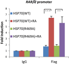
Fig. S6.
Wild-type or mutant HSP70 (R469A) binding with chromatin. HEK293T cells transfected with Flag-tagged wild-type or mutant HSP70 (R469A) were treated with or without RA (10−7 M) for 1 h before ChIP with IgG or anti-Flag antibody. Binding of HSP70 on RARβ2 gene promoter region was examined through qPCR (±SEM, **P < 0.01, ***P < 0.001).
Fig. S7.
Fig. S7.
The expression levels of PRMT4 and JMJD6 examined by RT-qPCR. The expression levels of PRMT4 (A) and JMJD6 (B) in samples described in Fig. 5 F and J, respectively, were examined through RT-qPCR.
Fig. 6.
Fig. 6.
HSP70 is required for the recruitment of TFIIH during RA-induced RARβ2 gene transcriptional activation. (A–D) Wild-type (hsp70.1+/+) MEFs were treated with or without RA (10−7 M) for 1 h and then subjected to ChIP with control IgG or RARα, RXRα (A); NCoR, p300 (B); MED6, MED1 (C); Pol II, TFIIB, TFIID (TBP) (D) antibody. Binding of these factors on the RARβ2 gene promoter region was examined through qPCR (±SEM, **P < 0.01, ***P < 0.001). (E) Wild-type (hsp70.1+/+) and hsp70-deficient (hsp70.1−/−) MEFs were treated with or without RA (10−7 M) for 1 h and then subjected to ChIP with control IgG or TFIIH (ERCC3) antibody. Binding of TFIIH (ERCC3) on the RARβ2 gene promoter region was examined through q-PCR (±SEM, ***P < 0.001).
Fig. S8.
Fig. S8.
Binding of receptors, corepressors, coactivators, mediators, and preinitiation complex in hsp70-deficient (hsp70.1−/−) MEFs. hsp70.1−/− MEFs were treated with or without RA (10−7 M) for 1 h, followed by ChIP with control IgG or RARα, RXRα (A); NCoR, p300 (B); MED6, MED1 (C); Pol II, TFIIB, TFIID (TBP) (D) antibody. Binding of these factors on RARβ2 gene promoter region was examined through qPCR (±SEM, *P < 0.05, **P < 0.01, ***P < 0.001).
Fig. 7.
Fig. 7.
HSP70 R469 methylation involved in TFIIH recruitment. (A) Cell lysates collected from HEK293T cells were subjected to IP with control IgG or anti-HSP70 antibody followed by IB with antibodies as indicated. (B) Cell lysates collected from HEK293T cells stably expressing control vector, Flag-tagged wild-type, or mutant HSP70 (R469A) were subjected to IP with anti-Flag antibody followed by IB with antibodies as indicated. (C) HEK293T cells were transfected with control siRNA or siRNA specifically against PRMT4 for 72 h and then treated with RA (10−7 M) for 1 h, followed by ChIP with TFIIH (ERCC3) antibody. Binding of TFIIH (ERCC3) on the RARβ2 gene promoter region was examined through qPCR. ChIP signals were presented as fold-induction by RA compared with control after normalization to input (±SEM, ***P < 0.001). (D) hsp70.1−/− MEFs were transfected with control vector, Flag-tagged wild-type or mutant HSP70 (R469A) for 48 h, and then treated with RA (10−7 M) for 1 h, followed by ChIP with TFIIH (ERCC3) antibody. Binding of TFIIH (ERCC3) on RARβ2 gene promoter region was examined through qPCR (±SEM, ***P < 0.001). (E) The expression of HSP70 vectors as described in D was examined by IB. (F) Model: HSP70 arginine methylation in RA-induced RARβ2 gene transcriptional activation. Upon RA stimulation, the binding of receptors (RARα and RXRα), mediator complex (such as MED1 and MED6), preinitiation complex (PIC, such as Pol II, TFIIA, TFIIB, TFIID, TFIIE, TFIIF, and TFIIH), HSP70, and PRMT4 were further increased on RARβ2 gene promoter, accompanied by the exchange of corepressor complexes (such as NCoR) and coactivator complexes (such as p300). During this process, PRMT4-mediated monomethylation of R469 in HSP70 was required for the recruitment of TFIIH and transcription initiation. For simplicity, other factors shown to be essential for RA-induced RARβ2 gene transcriptional activation were not included.
Fig. S9.
Fig. S9.
Generation of HEK293T cells stably expressing Flag-tagged wild-type or mutant HSP70 (R469A). HEK293T cells were transfected with Flag-tagged wild-type (Upper) or mutant HSP70 (R469A) (Lower) expression vector and then selected with puromycin (2.5 mg/mL). The resultant single colonies were harvested, separated by Bis-Tris NuPAGE gel and subjected to IB with anti-HSP70 antibody. Colonies with stably expressed exogenous HSP70 were highlighted in red. Colony number two of both wild-type and mutant HSP70 (R469A) were selected for further experiments.

References

    1. Strahl BD, Allis CD. The language of covalent histone modifications. Nature. 2000;403(6765):41–45. - PubMed
    1. Tan M, et al. Identification of 67 histone marks and histone lysine crotonylation as a new type of histone modification. Cell. 2011;146(6):1016–1028. - PMC - PubMed
    1. Byvoet P, Shepherd GR, Hardin JM, Noland BJ. The distribution and turnover of labeled methyl groups in histone fractions of cultured mammalian cells. Arch Biochem Biophys. 1972;148(2):558–567. - PubMed
    1. Murray K. The occurrence of epsilon-N-nethyl lysine in histones. Biochemistry. 1964;3:10–15. - PubMed
    1. Shi Y, et al. Histone demethylation mediated by the nuclear amine oxidase homolog LSD1. Cell. 2004;119(7):941–953. - PubMed

Publication types