. 2015 Jun;7(6):370-82.
doi: 10.18632/aging.100756.

Interstitial pericytes decrease in aged mouse kidneys

Affiliations

Interstitial pericytes decrease in aged mouse kidneys

Ania Stefanska et al. Aging (Albany NY). 2015 Jun.

Abstract

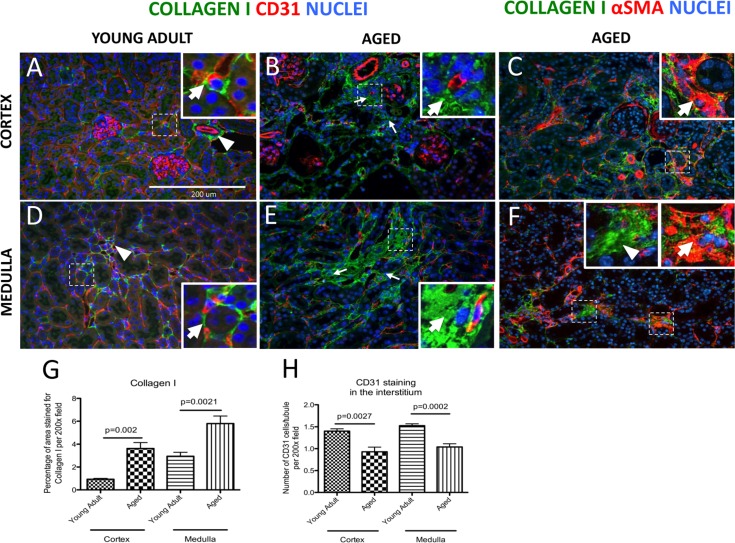
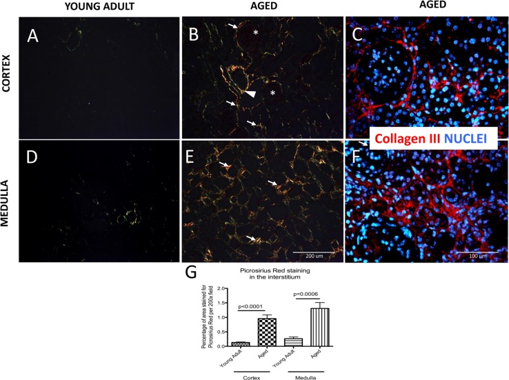
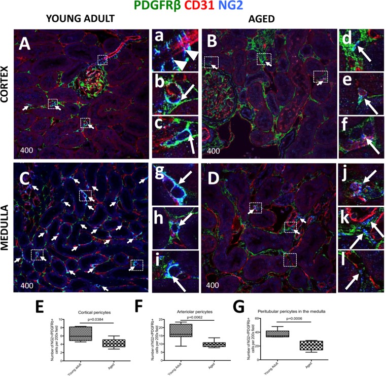
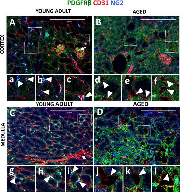
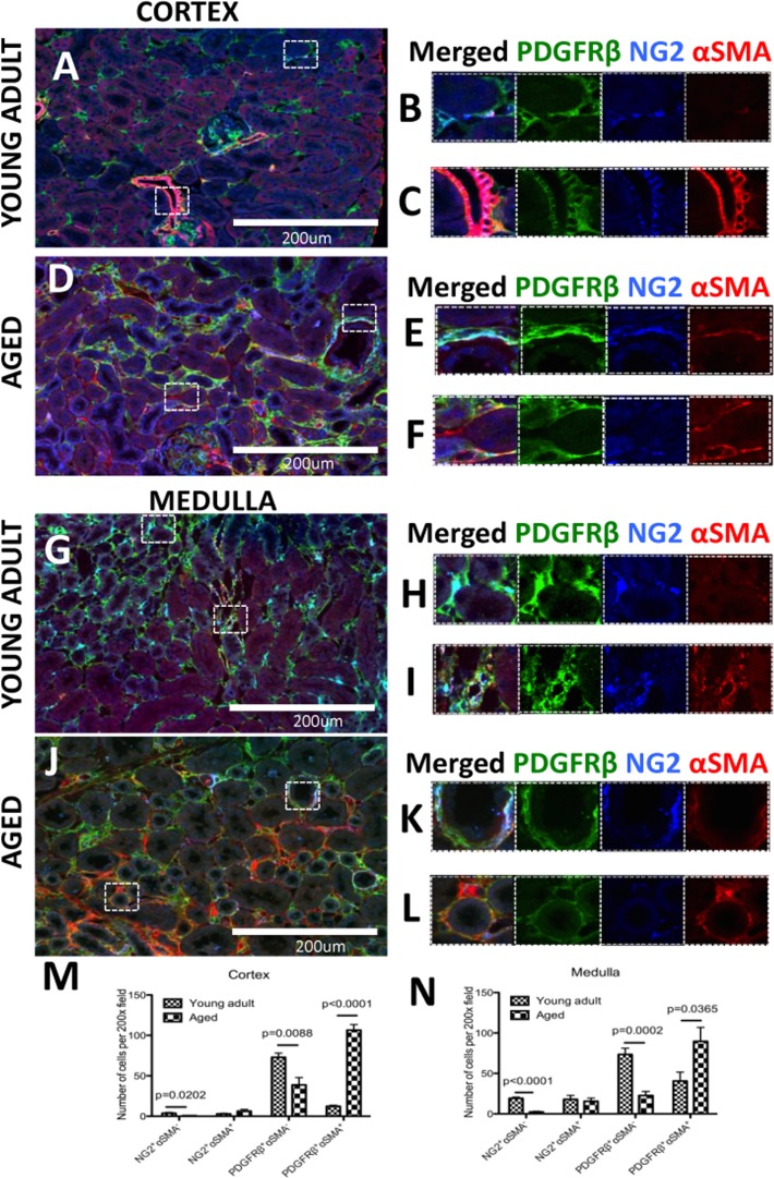
With increasing age, the kidney undergoes characteristic changes in the glomerular and tubulo-interstitial compartments, which are ultimately accompanied by reduced kidney function. Studies have shown age-related loss of peritubular vessels. Normal peritubular vessel tone, function and survival depend on neighboring pericytes. Pericyte detachment leads to vascular damage, which can be accompanied by their differentiation to fibroblasts and myofibroblasts, a state that favors matrix production. To better understand the fate of pericytes in the aged kidney, 27 month-old mice were studied. Compared to 3 month-old young adult mice, aged kidneys showed a substantial decrease in capillaries, identified by CD31 staining, in both cortex and medulla. This was accompanied by a marked decrease in surrounding NG2+ / PDGFRβ+ pericytes. This decrease was more pronounced in the medulla. Capillaries devoid of pericytes were typically dilated in aged mice. Aged kidneys were also characterized by interstitial fibrosis due to increased collagen-I and -III staining. This was accompanied by an increase in the number of pericytes that acquired a pro-fibrotic phenotype, identified by increased PDGFRβ+ / αSMA+ staining. These findings are consistent with the decline in kidney interstitial pericytes as a critical step in the development of changes to the peritubular vasculature with aging, and accompanying fibrosis.

Keywords: NG2; PDGFβ-receptor; endothelium; nephropathy; tubulo-interstitial fibrosis.


Conflict of interest statement

Conflict of interest statement

None of the authors have any financial or other conflicts of interest. The results presented in this paper have not been published previously in whole or part.

Figures

Figure 1
Figure 1. Increase in interstitial fibrosis and reduction of microvascular density in aged mice kidneys
Fibrosis was measured by staining for collagen I (green color). Double staining of αSMA (intracellular antigen) and collagen I was used to discern collagen I-expressing myofibroblasts from extracellular matrix proteins. Microvascular rarefaction was assessed by CD31 staining (red). DAPI staining identifies nuclei (blue). (A) Collagen I staining was faint and confined to the fibroblasts/pericytes in the cortex of young adult kidneys. The inset shows a high power image of a collagen I+ cell (green) encircling capillary (red) (arrow). Adventitial cells of renal arteries also stain for collagen I (arrowhead). (B) Collagen I staining was more abundant in the cortical interstitium of aged kidneys (arrows indicate examples) and was found outside the capillary walls (inset, arrow shows collagen I staining). CD31 staining was reduced in intensity. (C) Co-staining for collagen I and αSMA shows the presence of myofibroblasts (inset, arrow). (D) Collagen I staining in the medulla of young adult mice was present in vasa recta (arrowhead) and perivascular fibroblasts (inset, arrow). (E) In aged kidneys interstitial collagen I staining was increased (arrows indicate examples). The inset shows high power view of accumulation of collagen I staining (arrow). CD31 staining was decreased. (F) Co-staining of collagen I with αSMA confirms the presence of myofibroblasts (inset, arrow). Extracellular collagen I was also detected (inset, arrowhead). (G) Graph showing quantification of interstitial collagen I staining was significantly increased in both the cortex and the medulla. (H) Quantification of CD31 staining shows reduced vascular density per tubule in both the cortex and the medulla. Data are represented as mean ± SEM (n=6).
Figure 2
Figure 2. Accumulation of type I and III collagen in aged kidneys
Picrosirius Red staining of collagen I and III was examined by polarized light microscopy. Additionally, collagen III fibers are visualized with specific antibody (red color), nuclei are labeled with dapi (blue color). (A) Collagen fibers were sparse in the cortex of young adult kidneys. (B) Aged kidneys demonstrated interstitial collagen deposition (arrows indicate examples) in the peritubular and periglomerular area (glomeruli are indicated by asterix). Adventitia of arterial wall was enriched in collagen (arrowhead). (C) Collagen III staining confirms the location of interstitial and periglomerular fibrosis. (D) Picrosirius Red was barely detected in the medulla of young adult kidneys. (E) Picrosirius Red staining was markedly increased in the medulla of aged mice (arrows indicate examples) in the interstitium. (F) Collagen III staining confirms tubulointerstital fibrosis. (G) Graph of quantitation: Picrosirius Red staining significantly increased in the interstitium in both the cortex and the medulla of aged mice. Data are represented as mean ± SEM (n=6).
Figure 3
Figure 3. Decreased expression of kidney pericyte markers in aged mice
Pericytes were identified by double NG2+/PDGFRß+ staining (blue and green colors respectively), and their perivascular location was identified by endothelial CD31 staining (red color). Double positive cells (light blue color) were quantified using single channel images (marked here by the arrows). (A) In young adult kidney cortex, staining for pericyte markers was readily detected alongside afferent arterioles (a, arrowheads) and peritubular capillaries (examples indicated in the boxed regions b and c, arrows). (B) In aged kidney cortex, the number of cells staining for pericyte markers and lining interstitial peritubular capillaries was reduced. Pericytes surrounding the vessels show loss of contacts with endothelial cells (d, arrow), and CD31 staining intensity was reduced (e, f, arrows). (C) Kidney medulla of young adult mice showed abundant pericytes located along peritubular capillaries. The boxed regions show higher power images of pericytes tightly encircling peritubular capillaries (g, h, i, arrows). (D) In aged kidney medulla, there was a decline in number of pericytes surrounding peritubular capillaries. Pericyte staining intensity diminished (examples are shown in the boxed regions j, k, l arrows), and the vessel coverage was lacking (k,l, arrows). In aged mice compared to young adults, pericyte number was decreased in: (E) peritubular capillaries in the cortex, (F) pre-capillary arterioles, (G) peritubular capillaries in the medulla. Data are represented as mean ± SEM (n=6).
Figure 4
Figure 4. Capillary dilation in aged mice was associated with reduced pericyte coverage
Endothelial cells were marked by CD31 expression (red color). Pericytes were labelled by NG2+/PDGFRß+ staining (blue and green colors respectively). Double positive cells (light blue) were quantified using single channel images. (A) In young adult kidney cortex, peritubular capillaries form a regular pattern around renal tubules and inside the glomerular tuft (arrow). The boxed regions show higher power images of interstitial pericytes supporting peritubular capillaries (a, b, c, arrowheads). (B) In aged kidney cortex, endothelial cells become dilated, have decreased pericyte coverage (d, e, arrowheads) and some were completely isolated from tubules (f, arrowheads). (C) In young adult medulla, endothelial cells occupy peritubular spaces and vasa recta (arrow). Typically, few capillaries surround each tubule and pericyte, and are closely attached to the endothelial cells (g, h, i, arrowheads). (D) In aged kidney medulla, dilated peritubular capillaries were also present (j, k, arrowheads). In some instances, dilated capillaries appear in the areas of increased interstitial PDGFRß+ staining (l, arrowhead).
Figure 5
Figure 5. A subset of pericytes differentiate into myofibroblasts and increase in aged kidneys
Pericytes were identified by NG2+/PDGFRß+ staining (blue and green colors respectively). αSMA was used as myofibroblast marker (red color). (A) In the young adult kidney cortex, (B) most pericytes do not express αSMA. However in preglomerular arterioles (C) αSMA expression was present together with NG2/PDGFRß staining. (D) In aged kidney, αSMA increased in pericytes (E) and PDGFRß+ /NG2 cells (F). (G) Medulla of young adult kidney showed the presence αSMA in some peritubular capillaries (H) and contractile vasa recta cells (I). In aged mouse kidney, (J) there was accumulation of αSMA+ cells co-expressing PDGFRß and NG2 (K) or PDGFRß only (L). Quantification of αSMA expression in NG2+ and PDGFRß + cells showed that PDGFRß+ cells outnumber NG2+ cells in both young adult and aged kidneys. There was a dramatic increase in PDGFRß+αSMA+ cells and the numbers of αSMA-cells expressing either PDGFRß or NG2 were significantly lower in both (M) the cortex and (N) the medulla. Data are represented as mean ± SEM (n=6).

References

    1. Brown WW, Abrass IB, Oreopoulos DG. Introduction: aging and the kidney. Advances in renal replacement therapy. 2000;7:1–3. - PubMed
    1. Glassock RJ, Rule AD. The implications of anatomical and functional changes of the aging kidney: with an emphasis on the glomeruli. Kidney Int. 2012;82:270–277. - PMC - PubMed
    1. Abrass CK. The nature of chronic progressive nephropathy in aging rats. Advances in renal replacement therapy. 2000;7:4–10. - PubMed
    1. Abdel-Rahman EM, Okusa MD. Effects of aging on renal function and regenerative capacity. Nephron Clinical practice. 2014;127:15–20. - PubMed
    1. Schmitt R, Cantley LG. The impact of aging on kidney repair. American journal of physiology Renal physiology. 2008;294:F1265–1272. - PubMed

Publication types

Substances