. 2015 Jun 17;17(1):162.
doi: 10.1186/s13075-015-0682-6.

Autoantigen microarrays reveal autoantibodies associated with proliferative nephritis and active disease in pediatric systemic lupus erythematosus

Affiliations

Autoantigen microarrays reveal autoantibodies associated with proliferative nephritis and active disease in pediatric systemic lupus erythematosus

D James Haddon et al. Arthritis Res Ther. .

Abstract

Introduction: Pediatric systemic lupus erythematosus (pSLE) patients often initially present with more active and severe disease than adults, including a higher frequency of lupus nephritis. Specific autoantibodies, including anti-C1q, anti-DNA and anti-alpha-actinin, have been associated with kidney involvement in SLE, and DNA antibodies are capable of initiating early-stage lupus nephritis in severe combined immunodeficiency (SCID) mice. Over 100 different autoantibodies have been described in SLE patients, highlighting the need for comprehensive autoantibody profiling. Knowledge of the antibodies associated with pSLE and proliferative nephritis will increase the understanding of SLE pathogenesis, and may aid in monitoring patients for renal flare.

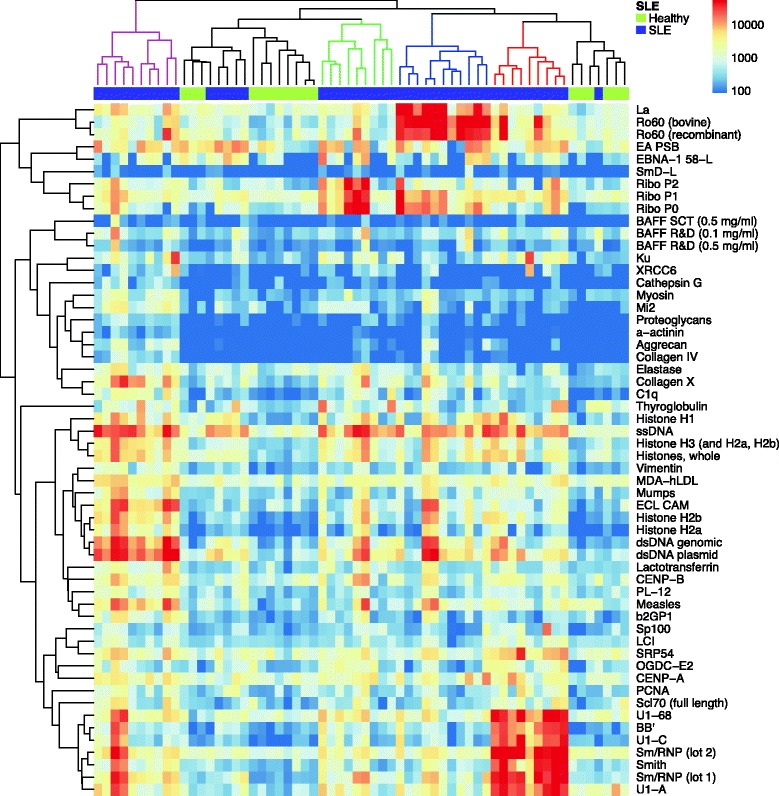
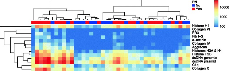
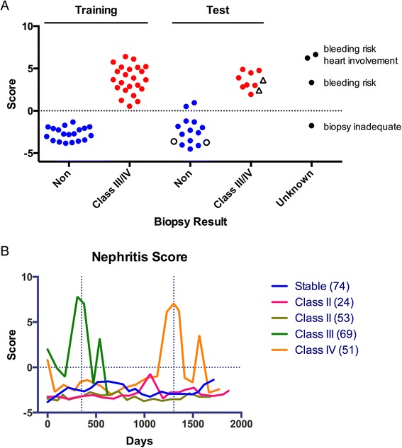
Methods: We used autoantigen microarrays composed of 140 recombinant or purified antigens to compare the serum autoantibody profiles of new-onset pSLE patients (n = 45) to healthy controls (n = 17). We also compared pSLE patients with biopsy-confirmed class III or IV proliferative nephritis (n = 23) and without significant renal involvement (n = 18). We performed ELISA with selected autoantigens to validate the microarray findings. We created a multiple logistic regression model, based on the ELISA and clinical information, to predict whether a patient had proliferative nephritis, and used a validation cohort (n = 23) and longitudinal samples (88 patient visits) to test its accuracy.

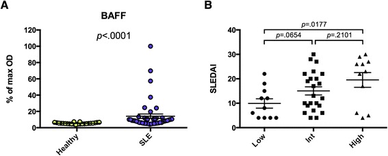
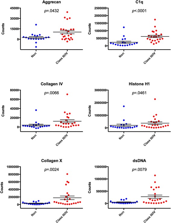
Results: Fifty autoantibodies were at significantly higher levels in the sera of pSLE patients compared to healthy controls, including anti-B cell-activating factor (BAFF). High levels of anti-BAFF were associated with active disease. Thirteen serum autoantibodies were present at significantly higher levels in pSLE patients with proliferative nephritis than those without, and we confirmed five autoantigens (dsDNA, C1q, collagens IV and X and aggrecan) by ELISA. Our model, based on ELISA measurements and clinical variables, correctly identified patients with proliferative nephritis with 91 % accuracy.

Conclusions: Autoantigen microarrays are an ideal platform for identifying autoantibodies associated with both pSLE and specific clinical manifestations of pSLE. Using multiple regression analysis to integrate autoantibody and clinical data permits accurate prediction of clinical manifestations with complex etiologies in pSLE.


Figures

Fig. 1
Fig. 1
Autoantigen microarrays identify autoantibodies associated with pediatric SLE. Serum samples from 45 new-onset pSLE patients and 17 age-matched healthy controls were used to probe autoantigen microarrays featuring 140 purified or recombinant autoantigens. Cy5-conjugated anti-human IgG antibodies were used as a secondary reagent, a fluorescent microarray scanner was used to image each microarray, and the feature MFIs were used to quantify binding. Significance analysis of microarrays (SAM) was used to identify significant differences in IgG reactivity to the autoantigens between pSLE patients and controls (q-value <0.001, fold change >2). A hierarchically clustered heatmap (complete linkage, unsupervised) of the significant autoantigens is shown, with pSLE patients indicated on the top bar in purple and healthy controls in green. Patient clusters are colored red, blue green and magenta in the phylogenetic tree. IgG immunoglobulin G, MFI median fluorescence intensity, pSLE pediatric systemic lupus erythematosus, SLE systemic lupus erythematosus
Fig. 2
Fig. 2
Anti-BAFF is present in a subset of pediatric SLE patients with active disease. a Recombinant BAFF was used to coat 96-well ELISA plates, and sera from 45 pSLE patients and 24 age-matched healthy controls were used to probe them in triplicate. HRP-conjugated goat anti-human IgG (heavy and light chain) was used as a secondary reagent. After development with TMB substrate, absorbance was measured at 450 nm, and each sample is shown as a percentage of the maximum absorbance. b The pSLE patients were divided into groups based on their serum reactivity to BAFF. The modified SELENA-SLEDAI is shown for the lowest quartile (Low, <5.8), middle two quartiles (Int), and highest quartile (High, >11.0). Mann–Whitney tests were used to compare reactivity between groups (bars show mean ± SEM). BAFF B cell-activating factor, ELISA enzyme-linked immunosorbent assay, IgG immunoglobulin G, HRP horseradish peroxidase, pSLE pediatric systemic lupus erythematosus, SELENA-SLEDAI Safety of Estrogen in Lupus Erythematosus National Assessment-SLE disease activity index, SLE systemic lupus erythematosus, TMB 3,3′,5,5′-tetramethylbenzidine
Fig. 3
Fig. 3
Autoantigen microarrays identify multiple autoantibodies associated with proliferative nephritis in pSLE. Serum samples from 41 new-onset pSLE patients were used to probe autoantigen microarrays featuring 140 purified or recombinant autoantigens. Anti-human IgG antibodies conjugated with Cy5 were used as a secondary reagent, a fluorescent microarray scanner was used to image each microarray, and the feature MFIs were used to quantify binding. Significance analysis of microarrays (SAM) was used to identify significant differences in IgG reactivity to the autoantigens between patients with biopsy-proven class III/IV nephritis and patients without significant evidence of nephritis (q-value <0.06, fold change >2). A hierarchically clustered heatmap of the significant antigens is shown. Patients with biopsy-proven proliferative nephritis are indicated on the top bar in red, while patients without nephritis are shown in blue. IgG immunoglobulin G, MFI median fluorescence intensity, pSLE pediatric systemic lupus erythematosus, PN proliferative nephritis
Fig. 4
Fig. 4
ELISA validation of candidate autoantigens identified by microarray. Significance analysis of microarrays (SAM) identified 13 autoantigens as having significantly different IgG reactivity between pSLE patients with and without proliferative nephritis, of which seven were selected for further validation using indirect serum ELISA. Purified or recombinant forms of the autoantigens shown above were used to coat 96-well ELISA plates. Serum from 42 pSLE patients was used to probe them in duplicate, and europium-labeled anti-human IgG (Fcγ specific) was used as a secondary reagent. Time-resolved fluorescent counts for six of the seven selected autoantigens (minus signal from BSA-coated wells) are shown above. Mann–Whitney tests were used to compare reactivity between groups (bars show mean ± SEM). BSA bovine serum albumin, ELISA enzyme-linked immunosorbent assay, IgG immunoglobulin G, pSLE pediatric systemic lupus erythematosus
Fig. 5
Fig. 5
Identification of a pediatric SLE proliferative nephritis predictive signature. a Least absolute shrinkage and selection operator (LASSO) was used on a training set of ELISA and clinical measurements from 41 new-onset pSLE patient samples as a variable selection and linear regression method to construct a predictive model of proliferative nephritis. A separate test set of 23 new-onset pSLE patient samples was used to evaluate the performance of the model, and the nephritis scores obtained for each patient with the model are shown. Unfilled black circles indicate patients who were biopsied and found to be class II, and unfilled black triangles indicate class V nephritis patients. The model was also used to calculate nephritis scores for samples from new-onset pSLE patients who were suspected to have nephritis, but were not confirmed by biopsy (unknown). b Five pSLE patients from the test set were selected for longitudinal analysis. ELISA was used to determine each serum sample’s anti-dsDNA and anti-C1q IgG levels, and a chart review was performed to collect all applicable clinical data. The LASSO model based on the training set was used to calculate nephritis scores for each patient at each time point. Vertical dashed lines indicate when biopsies were performed, and the horizontal dashed line indicates the nephritis score cutoff. ELISA enzyme-linked immunosorbent assay, IgG immunoglobulin G, pSLE pediatric systemic lupus erythematosus, SLE systemic lupus erythematosus

References

    1. Arbuckle MR, McClain MT, Rubertone MV, Scofield RH, Dennis GJ, James JA, et al. Development of autoantibodies before the clinical onset of systemic lupus erythematosus. N Engl J Med. 2003;349:1526–33. doi: 10.1056/NEJMoa021933. - DOI - PubMed
    1. Sherer Y, Gorstein A, Fritzler MJ, Shoenfeld Y. Autoantibody explosion in systemic lupus erythematosus: more than 100 different antibodies found in SLE patients. Semin Arthritis Rheum. 2004;34:501–37. doi: 10.1016/j.semarthrit.2004.07.002. - DOI - PubMed
    1. ter Borg EJ, Horst G, Hummel EJ, Limburg PC, Kallenberg CG. Measurement of increases in anti-double-stranded DNA antibody levels as a predictor of disease exacerbation in systemic lupus erythematosus. A long-term, prospective study. Arthritis Rheum. 1990;33:634–43. doi: 10.1002/art.1780330505. - DOI - PubMed
    1. Ehrenstein MR, Katz DR, Griffiths MH, Papadaki L, Winkler TH, Kalden JR, et al. Human IgG anti-DNA antibodies deposit in kidneys and induce proteinuria in SCID mice. Kidney Int. 1995;48:705–11. doi: 10.1038/ki.1995.341. - DOI - PubMed
    1. Mina R, Brunner HI. Pediatric lupus–are there differences in presentation, genetics, response to therapy, and damage accrual compared with adult lupus? Rheum Dis Clin North Am. 2010;36:53–80. doi: 10.1016/j.rdc.2009.12.012. - DOI - PMC - PubMed

Publication types